Get Well Soon, Titanosuchid
Christmas was not particularly kind for one titanosuchid. Published on the day many were receiving gifts and well wishes, this Permian reptile was given the bad news that it was suffering from a bout of osteomyelitis, a form of bone infection, at the time of its death. I have been working on a little side project involving palaeopathologies of late, the lovechild of clinical and veterinary medicine, visualisation techniques and vertebrate evolution. Whilst my focus has been on the ailments that plagued the dinosaurs, this diagnosis in a completely unrelated reptile serves as a timely reminder that injury and disease can be found throughout the animal kingdom, regardless of clade or geological age.
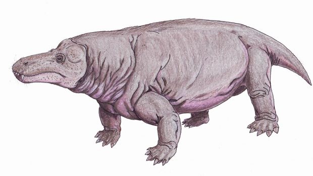
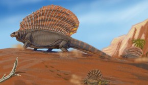
The titanosuchid in question is Jonkeria para, a South African therapsid, a clade of reptiles whose cousins would go on to spawn us mammals. As such, its suchian affinities are somewhat of a misnomer; whilst they were relatively large, as the name implies, they are of no relation to crocodiles and their kin by any stretch of the imagination. Jonkeria comes in at roughly 4-meters in length, with an elongate snout and stout little legs. Its role in the 265 million year old Karoo Basin ecosystem is somewhat unclear, however, with most in the field happy to call it an omnivore.
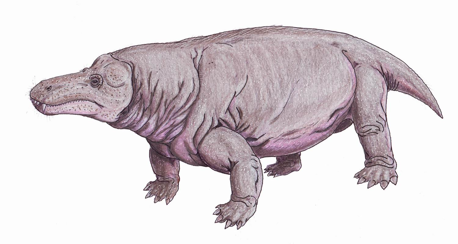
Jonkeria, a large therapsid that suffered from an infected bite wound to its thigh. This went on to affect the bone causing a condition known as osteomyelitis. Credit: ДиБгд, wikicommons.
Osteomyelitis is a general name for bone infection, and is contracted by various means. Infectious agents (usually bacteria, and in rare cases fungi) in the blood stream or adjacent organ systems, for instance, can subsequently invade bone tissue, whilst pathogens can de directly introduced following trauma to the bone, from a fracture or bite wound, for example. The latter is by far the most common, and thanks to the fact that bone reacts similarly to infection across vertebrates, it is possible to diagnose osteomyelitic bone from animals that have since turned to stone. Indeed, the presence of a bone reaction is one of the key ways palaeopathologists are able to diagnose sickness in ancient organisms, being clearly differentiated from damage caused during the fossilisation process.
The authors of the study best describe the etiology of the disease, explaining the struggle between the host’s immune system response and that of the invading pathogen:
“This study describes the non-granulomatous variety, wherein enzymes released by infectious agents erode the localised bone area. This results in holes or pockets. The body’s response is to attempt to wall off the infection by producing a surrounding reaction of new bone growth. As the infection continues, it erodes or dissolves through some of the new bone, such that there is a ‘contest’ as to which will be more effective. If the new bone is more effective, the infection becomes walled off. If the infection is more effective, it permeates the bone”
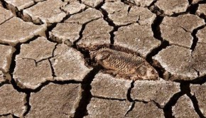
Two tooth punctures can be seen on the Jonkeria femur, a couple of sub circular depressions near to where it would articulate with the hip. Anteosaurus appeared the likely candidate, a half-tonne predatory therapsid with a beautiful set of canines. Yet the presence of infection is not immediately obvious. Some dinosaur specimens, for instance, show clear signs of infection thanks to the grossly enlarged nature of the bone; specimen AMNH 5207, a hadrosaur humerus, is known as the “sickest fossil bone” thanks to a severe fracture and subsequent misalignment, followed by modification of the bone surface as a result of an invasion by infectious agents. Furthermore, the Jonkeria femur was significantly weathered, highlighting some of the issues that plague palaeopathologists when it comes to identifying ancient diseases. Postmortem breaks, erosion and alterations to shape and texture can all have the appearance of sickness or injury, underlining the need for multiple or powerful visualisation techniques (preferably both) to accurately diagnose conditions.
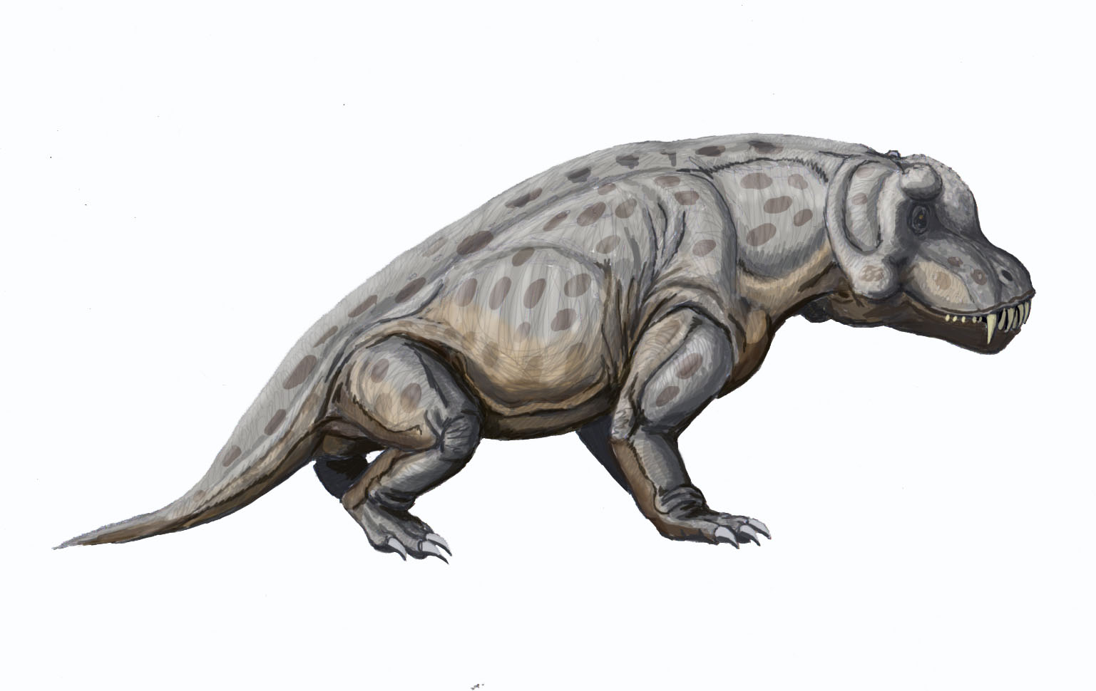
Anteosaurus, a large predatory therapsid from the Permian of South Africa, thought to be the author of the tooth-marked Jonkeria femur. Credit: ДиБгд, wikicommons.
As such, thin sections of bone from the Jonkeria specimen showed the deviant nature of its histology, possessing “unusual radial bony spicules extending from the periosteal surface to the medullary cavity”. The periosteum is the outside surface of bone, whereas the medullary cavity is the central cavity where marrow is stored, and this aberrant morphology is typical of a pathological response. Increased blood flow to the area was also noted, and next to the perisoteal reaction were little cavities synonymous with abscesses. The subadult nature of this Jonkeria individual also fits well with ideas that some predators may preferentially target substandard prey; why hunt adult individuals when you can go for an easier, more cost-effective meal? The length of time the animal lived with the infected, bite-marked bone was not ascertained, nor were its impact on its locomotion. It might be tempting to suggest, for instance, that the lack of exaggerated bone growth might suggest this animal died relatively quickly after its encounter with the predator. However, at this stage such comments are more conjecture than accurate science.
Whilst bone reaction to disease and injury is generally conservative across vertebrates, the rate of healing in ancient organisms is less well understood. Mammals heal significantly faster than reptiles, and the placement of this therapsid as a distant relative to modern day mammals raises interesting questions about their metabolism and subsequent immune system response: did they heal more like mammals or reptiles? The presence of fast growing fibrolamellar bone and Haversian systems (canals containing blood vessels and nerves) have been often thought to be traits of the ‘warm-blooded’ (forgive the terminology), suggesting Jonkeria possessed similar attributes, yet there is evidence these are not tied to any particular thermoregulatory profile*. Answering such questions is beyond the scope of this article, but leaves us with an interesting conundrum to ponder over as we recover from the festivities.
*For interested readers, The Complete Dinosaur (Eds: Brett-Surman, Holtz and Farlow) has several excellent chapters that focus on the evidence and caveats for the inference of thermoregulatory profiles in dinosaurs, some of which is applicable to ancient reptiles such as therapsids.
Related Posts
Episode 121/122: Dietary ecology of Smilodon fatalis →
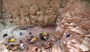
Episode 59: Chemnitz petrified forest →
Episode 49: Synapsids →