Episode 75: Laser-Stimulated Fluorescence
Palaeontology is a constantly evolving field; when new methods and techniques are invented, they allow us to revisit old fossils and test our previous observations and hypotheses. Recently, an exciting new method called ‘Laser-Simulated Fluorescence’ (LSF) has been gaining popularity in palaeontology and we speak to its inventor Tom Kaye, Foundation for Scientific Advancement, during a visit to the University of Bristol, alongside Dr Michael Pittman, Research Assistant Professor, The University of Hong Kong, China.
In this episode, we hear all about how LSF is allowing fossils to be seen under a completely new light. We discuss how the fluorescence is produced, how it’s currently being used and what possible applications it might have in future.
Podcast: Download (53.9MB)
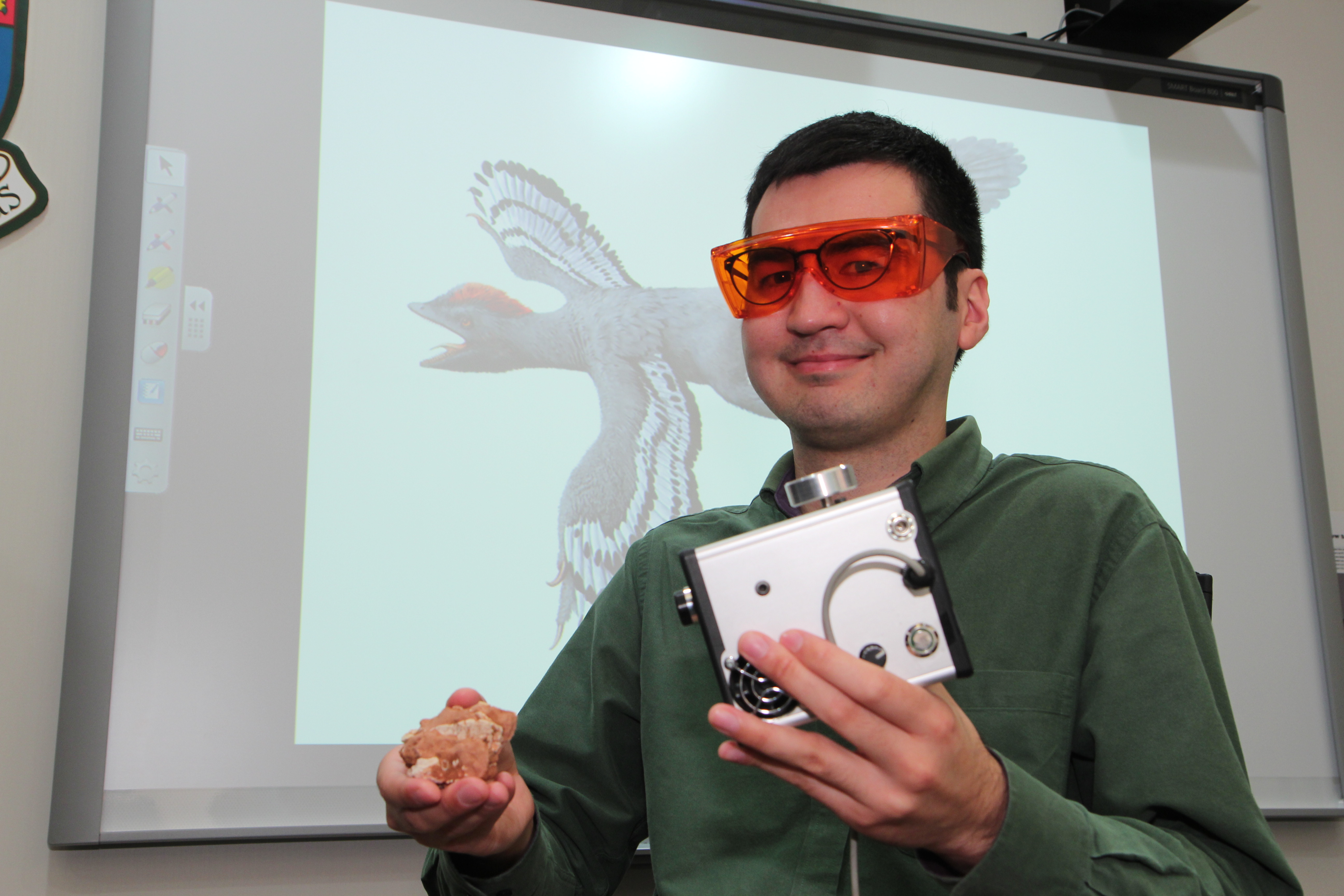
Dr Pittman and the laser-stimulated fluorescence device. (It’s about the size of walkman/ quarter a gramophone)
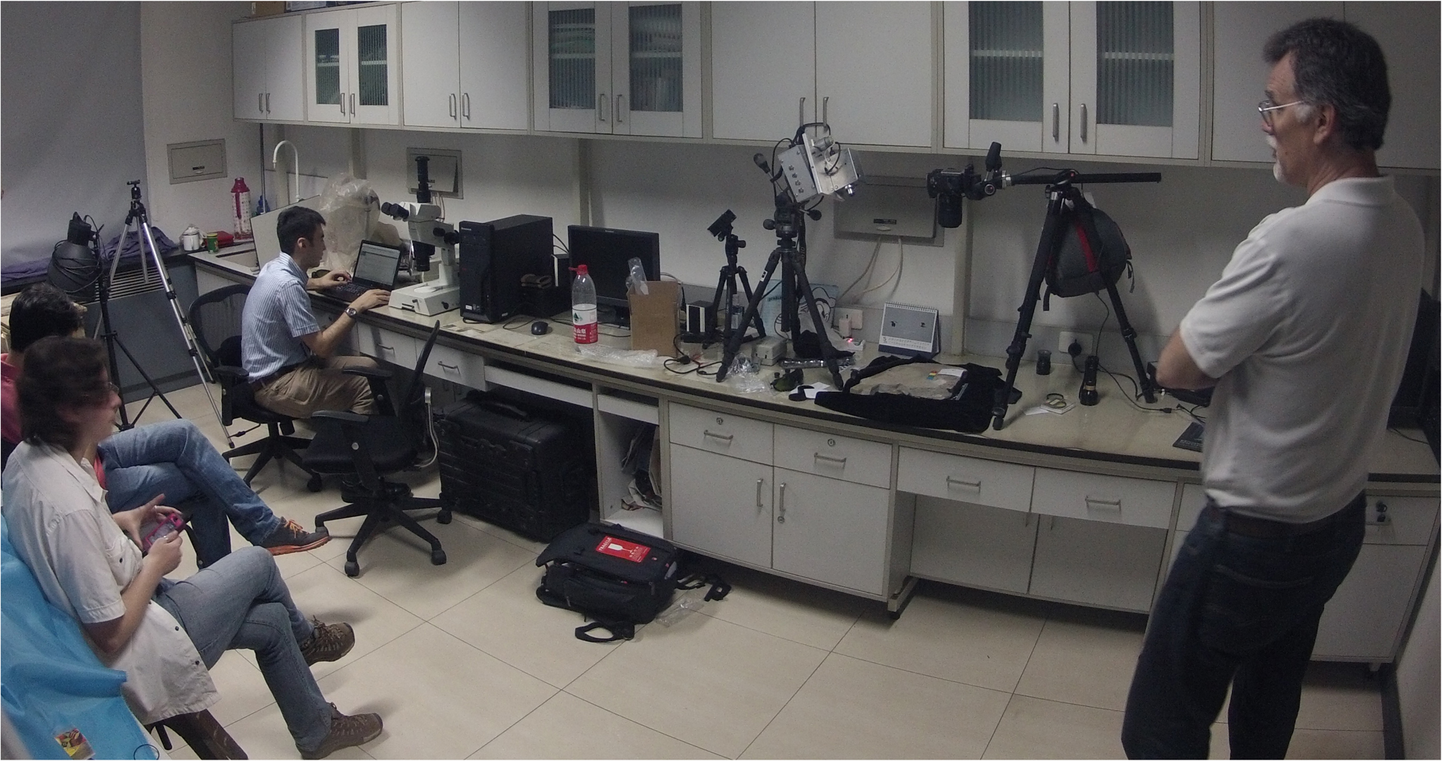
Dr Pittman and LSF inventor, Tom Kaye (right), have been employing the new method in locations all around the world, including this data collection trip to Beijing, China.
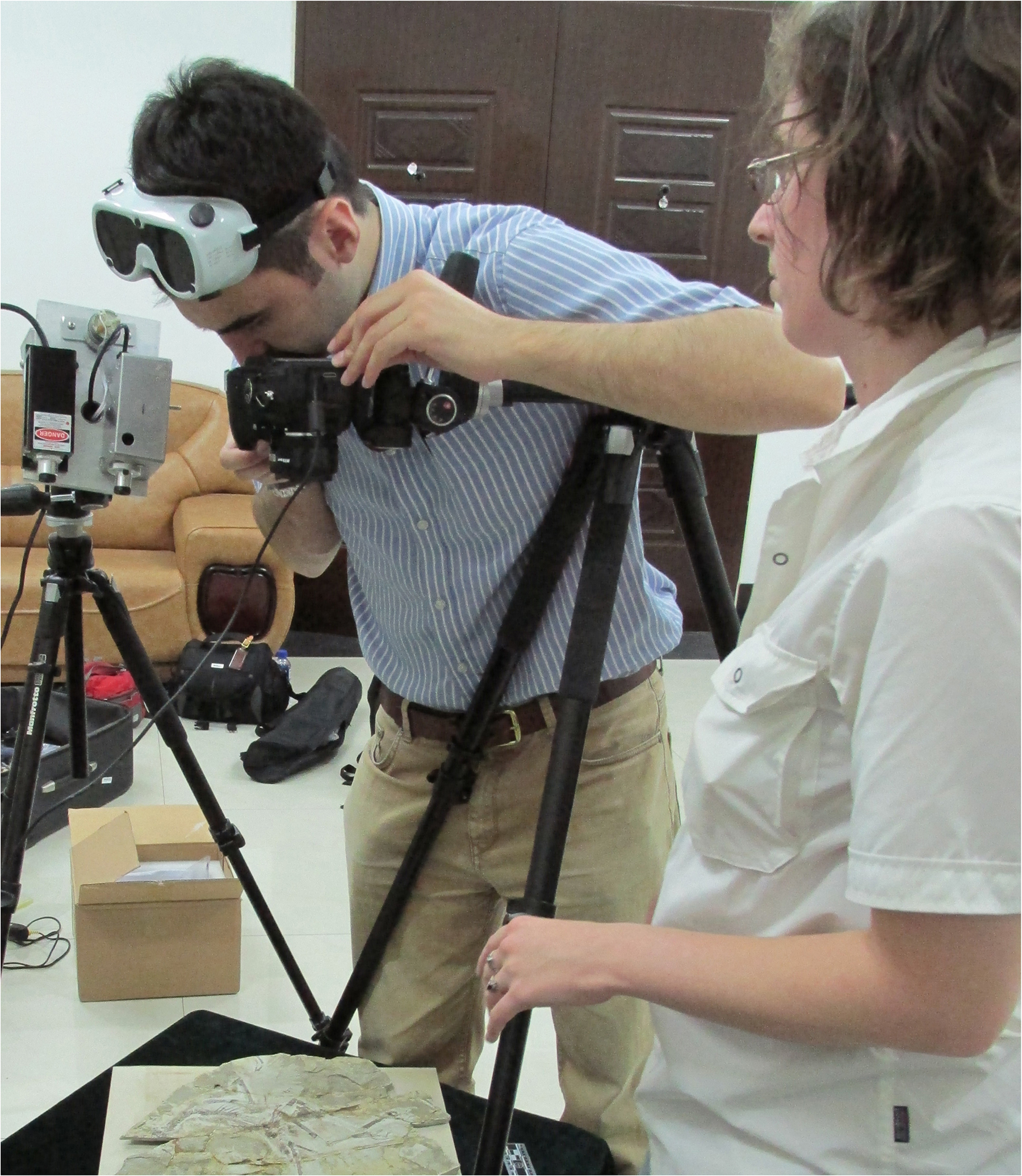
The LSF technology can be quickly set up and used to scan fossils. Also pictured is Dr Amanda Falk, Assistant Professor of Biology at Centre College and also the Palaeo After Dark podcast.
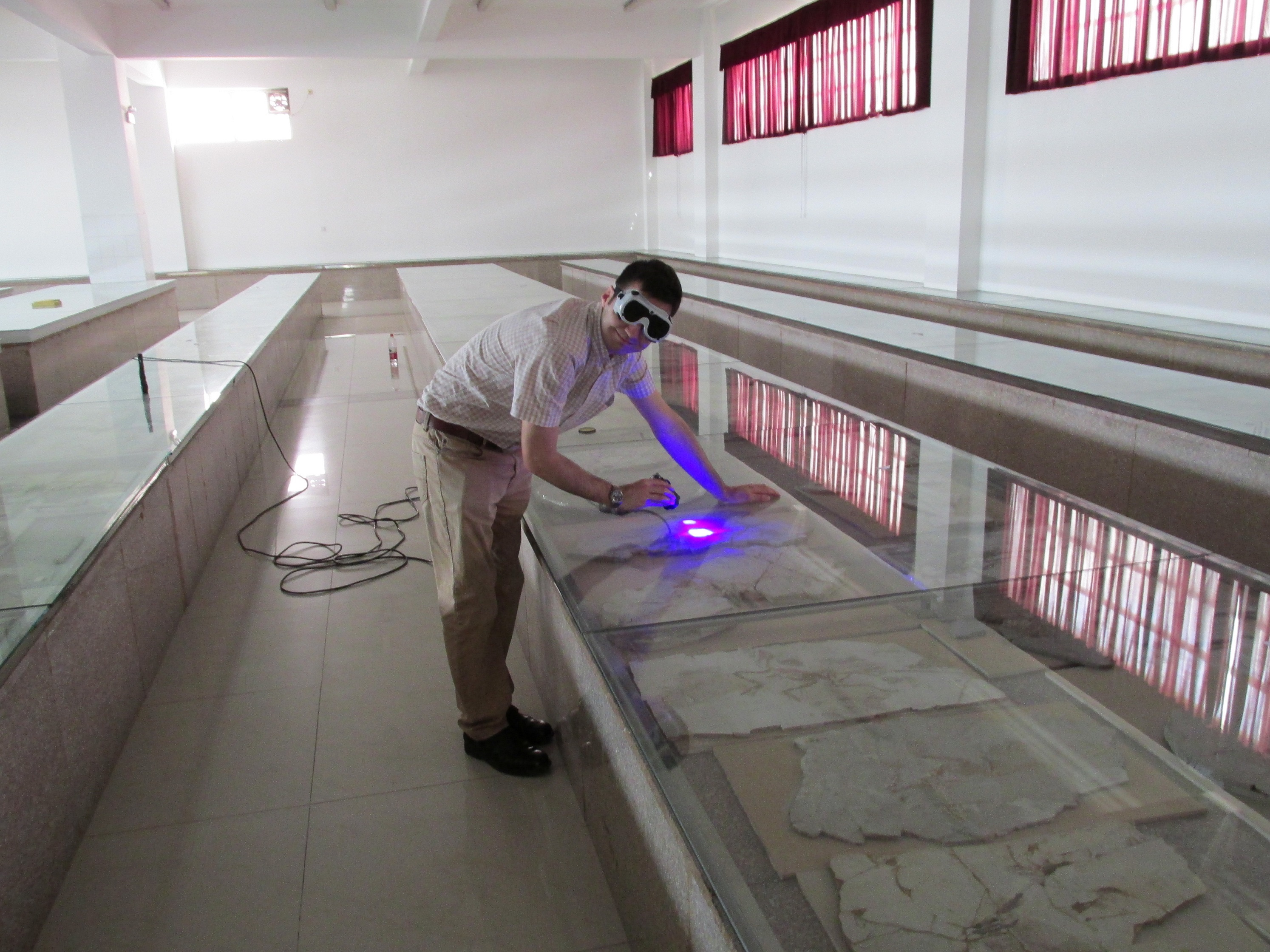
The laser is also handheld and can be used to rapidly scan multiple specimens with minimal disturbance. Any specimens of interest can then be removed and examined more thoroughly.
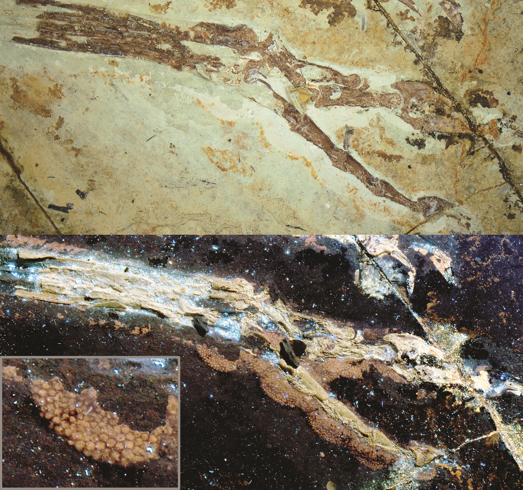
The LSF method allows for remarkable details to be revealed that would otherwise be invisible under normal light. Image: The foot of the bird-like feathered dinosaur Anchiornis. Top image shows the foot under normal light. Bottom image shows the foot under laser light. Inset image shows the scaly details of a single footpad. The footpad scales are preserved, but only visible under laser-stimulated fluorescence. Credit: Wang XL, Pittman M et al. 2017.
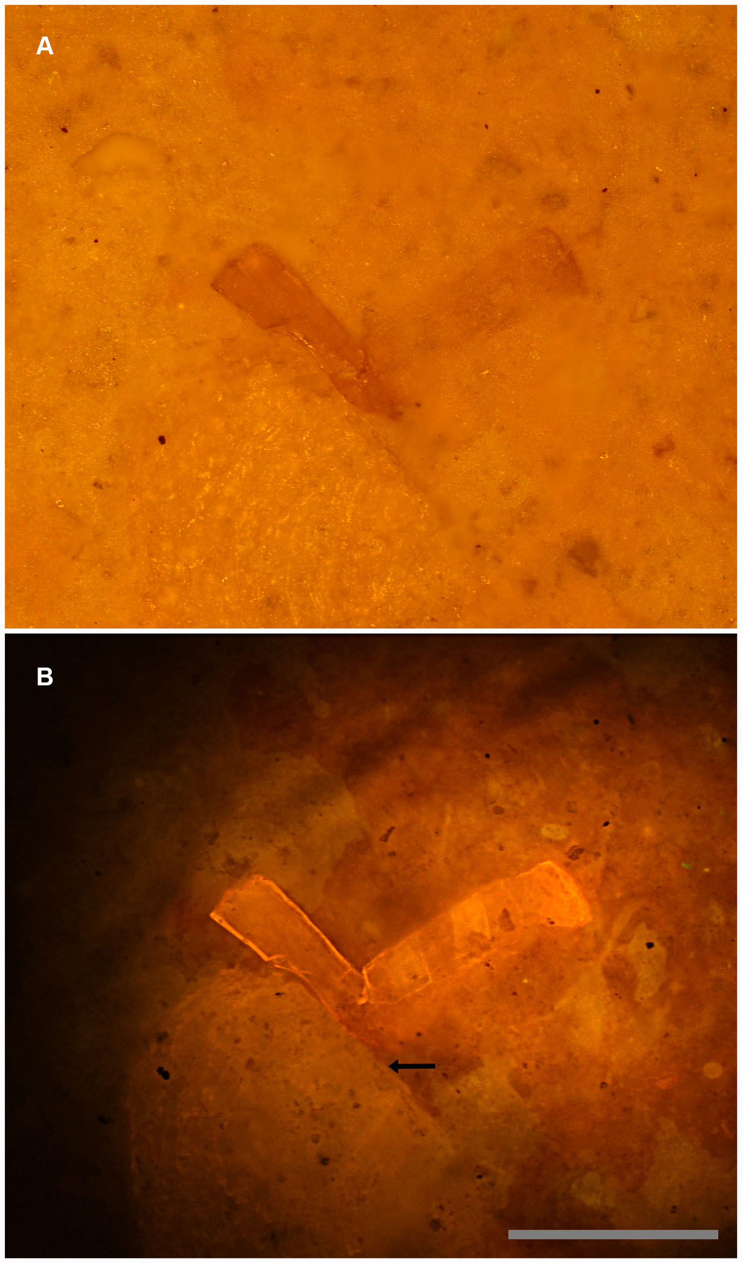
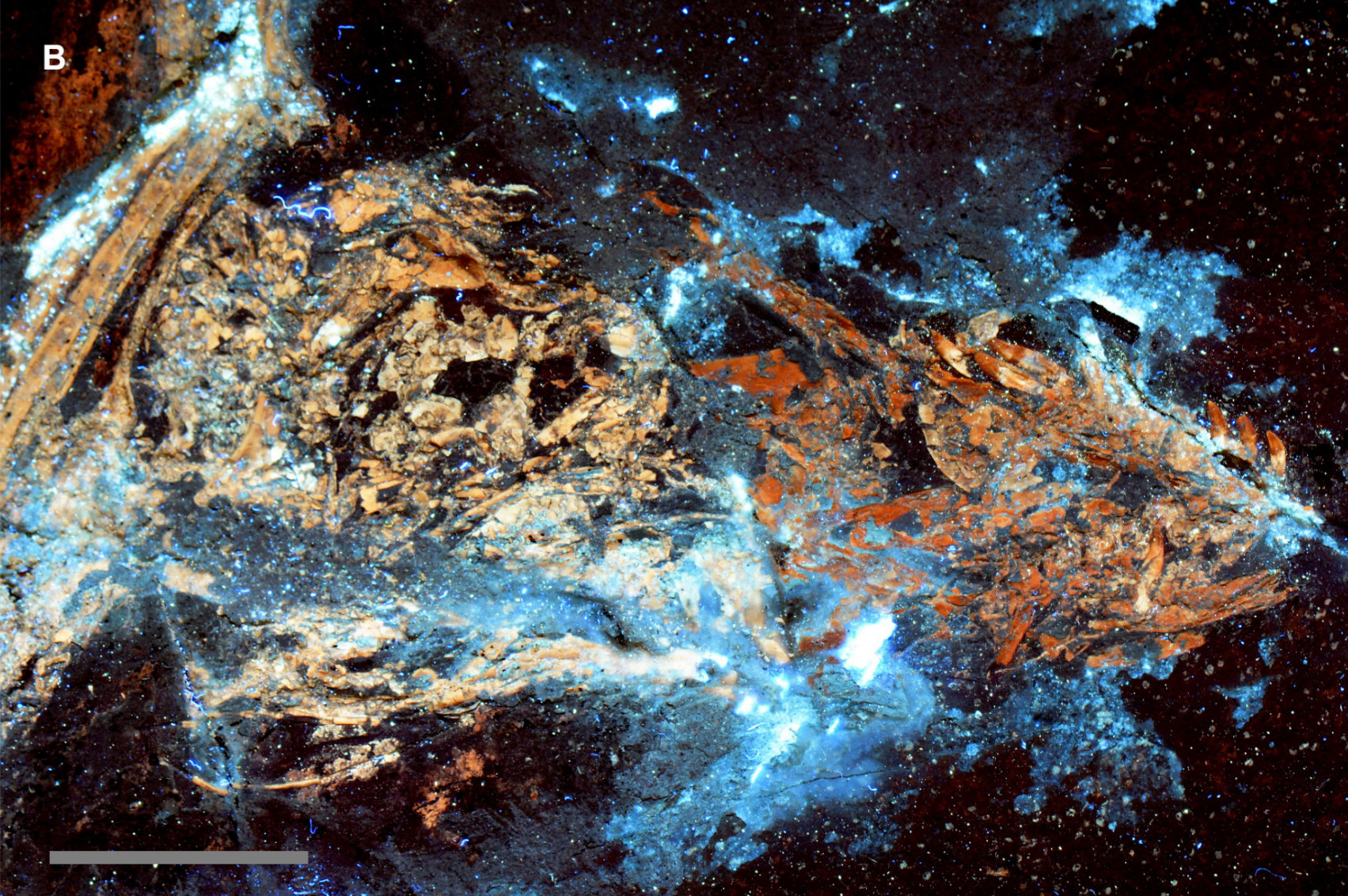
It’s not just superficial details either; the LSF can penetrate a short distance to reveal subsurface information. Image: A, White light photograph shows the bone fragment on the right entombed within matrix. B, Specimen under laser fluorescence. Photograph shows a high level of detail invisible under white light. Note that another much larger fragment also becomes visible (arrow). Scale bar 0.5 mm.
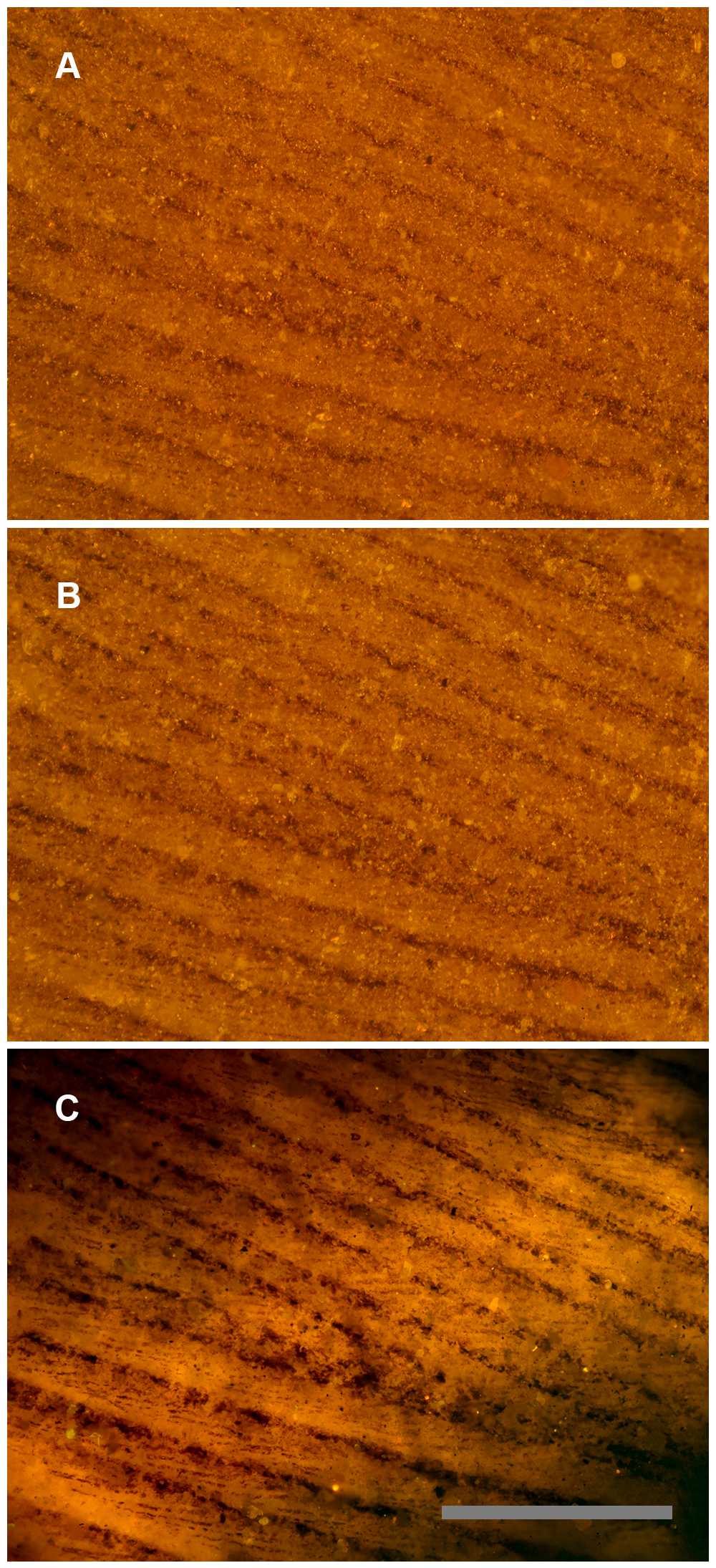
Comparison of different lighting methods, can reveal a wealth of additional information that our eyes cannot normally perceive. Image: A ~50 million year old bird feather from the Green River Formation, USA under different lighting conditions: A, White light, only barbs are visible. B, Polarized light, some traces of barbules. C, Laser-stimulated fluorescence of matrix behind the feather backlights the fossil, revealing the barbules in detail. Scale bar 0.5 mm.

This is not only useful for revealing new anatomical information, but can also be used to reinterpret fossils. In this instance, it was used to help determine whether the skull from a specimen of the gliding carnivorous dinosaur Microraptor was a composite or not. Image: The skull of specimen IVPP V13320 under white light conditions shows subtle colour differences in the bone across a break in the rock slab – darker bone proximally and lighter bone distally. Scale bar 1 cm.
Under laser light stimulation, the bone fluoresces with the same colour pattern observed under white light conditions indicating that the colour differences relate to differences in the fossil’s mineral composition. This indicates that the skull is a composite specimen, but it is also possible – but less likely – that the pattern observed reflects variability in the conditions under which the animal was deposited and eventually transformed into rock. Scale bar 1 cm.
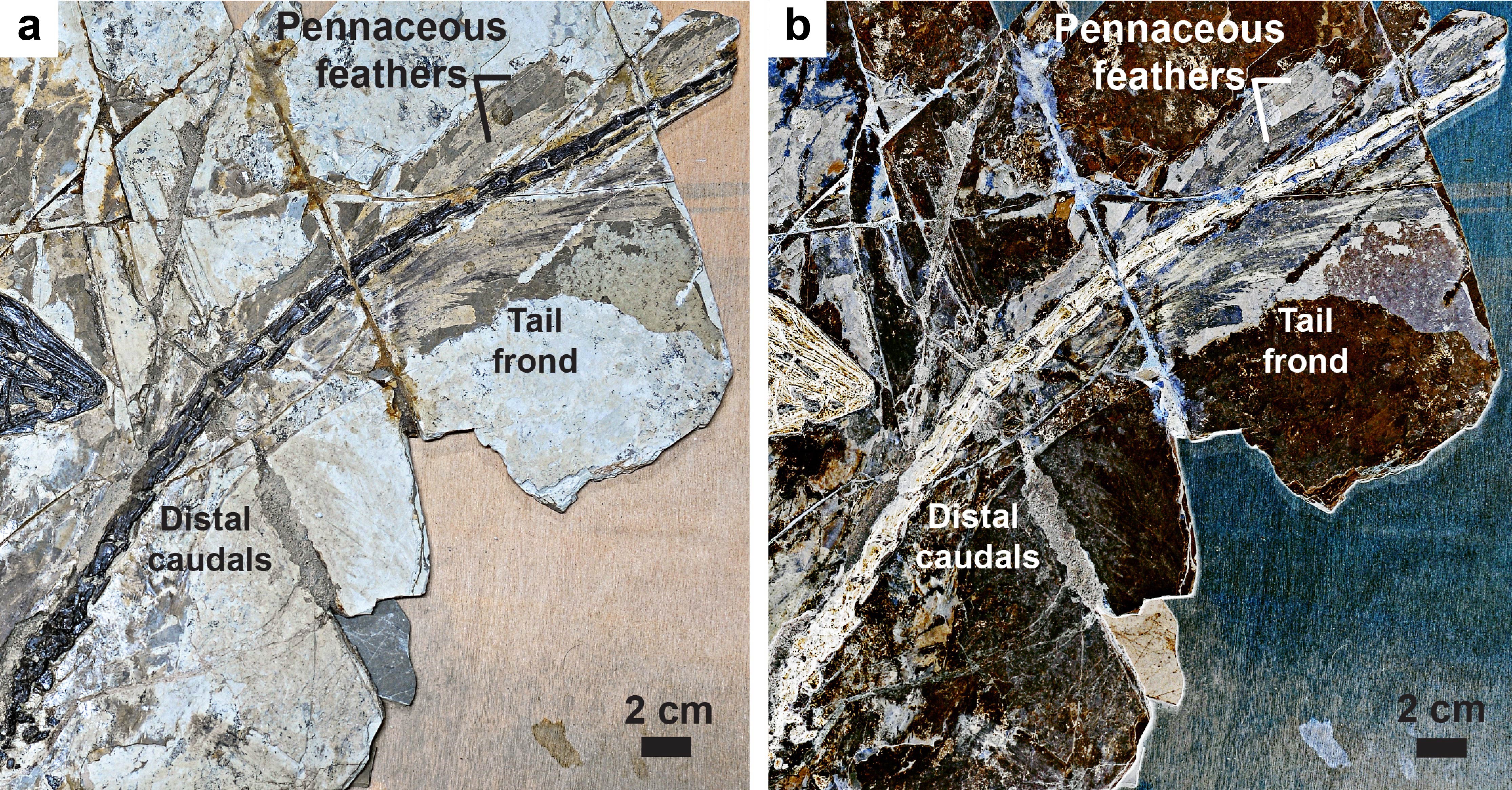
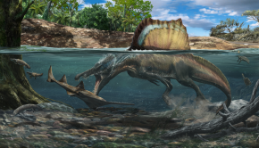
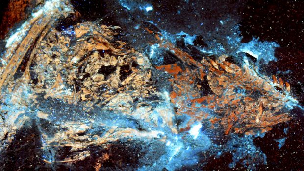
In research published today (at time of podcast release), the LSF method has been used to reveal the presence of flight-associated asymmetric feathers in a new species of dinosaur, extending the oldes record of these feathers to the closest common ancestor of birds and bird-like dinosaurs. Image: The tail frond of the troodontid Jianianhualong tengi DLXH 1218. (a) Photograph, and (b) LSF image of the fossil tail frond. Scale is 2cm. Credit. Xu, Currie, Pittman et. al. 2017.
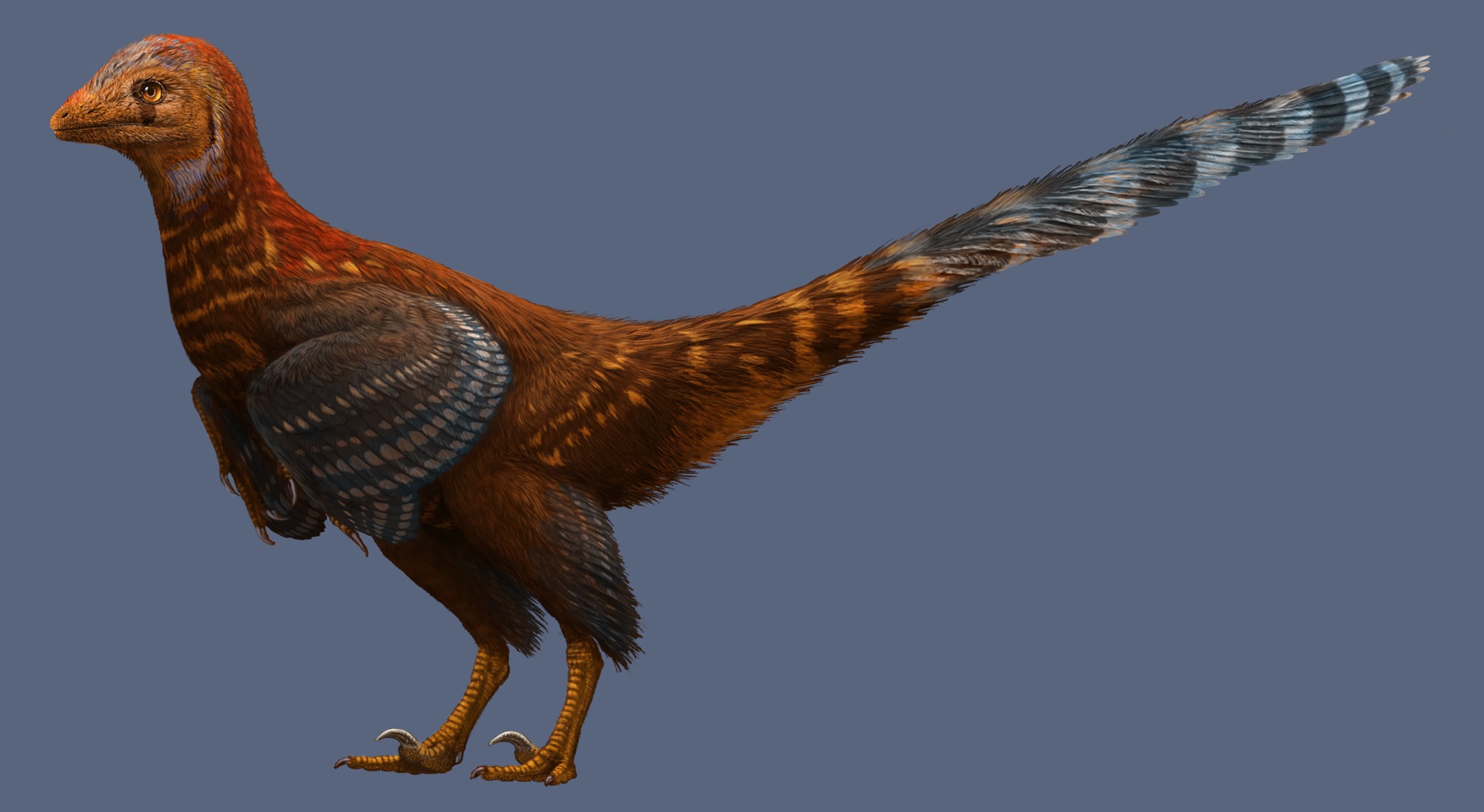
Such research demonstrates the clear benefit of using LSF to analyse fossils. Expect to see much wider use of this technique in future! Image: Life reconstruction of the asymmetrically feathered troodontid Jianianhualong tengi DLXH 1218. Credit: Julius T. Csotonyi 2017 / Xu, Currie, Pittman et. al. 2017.
All images courtesy of Dr Pittman unless otherwise stated.
Related Posts
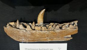
200 Years of Dinosaurs →
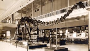
Episode 157: The Carnegie Diplodocus →
Life On Our Planet →