Feeding and the digestive system in early animals
Two new studies lead by University of Bristol palaeontologist Dr. Imran Rahman (Episode 28 – From worms to stars) are helping to explain feeding and the digestive system in some early animals. His work focuses on early echinoderms, which include modern animals like sea-stars and sea urchins, and these fossil organisms are related to these extant ones.
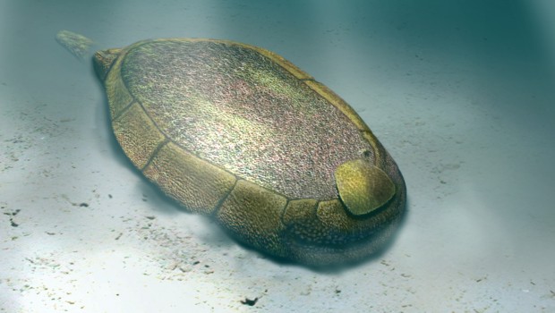
The first study (published in Proceedings of the Royal Society B) looks at a 510 million year old fossil found in Spain, and looked at reconstructing the feeding mode of this animal. The animal, Protocinctus mansillaensis, lived in the water and two different feeding methods had previously been debated: either it was an active filter feeder, or it passively waited for water and food to enter its mouth. The team used computer simulations to look at different feeding methods, and determined that it was an active pharyngeal filter feeder. This means that it used it’s internal gill slits of the pharynx to actively draw in water, trapping suspended food particles. By looking at the optimal position of Protocinctus in a current, they determined that the best way to reduce drag and lift would be to direct itself so the mouth was downstream, thereby making it extremely unlikely that it was a passive feeder since little food would actively end up in its mouth. The significance of this find was summarised by Dr. Rahman: “Humans and other vertebrates (animals with backbones) are part of a major group known as deuterostomes, which also includes invertebrates such as sea urchins, starfish and acorn worms. It has been very difficult to work out what the ancestor of all these groups looked like and how it fed because the modern forms are so different from one another. However, by studying one of the earliest fossil echinoderms with the aid of sophisticated methods we have been able to learn more about our ancient ancestry.”
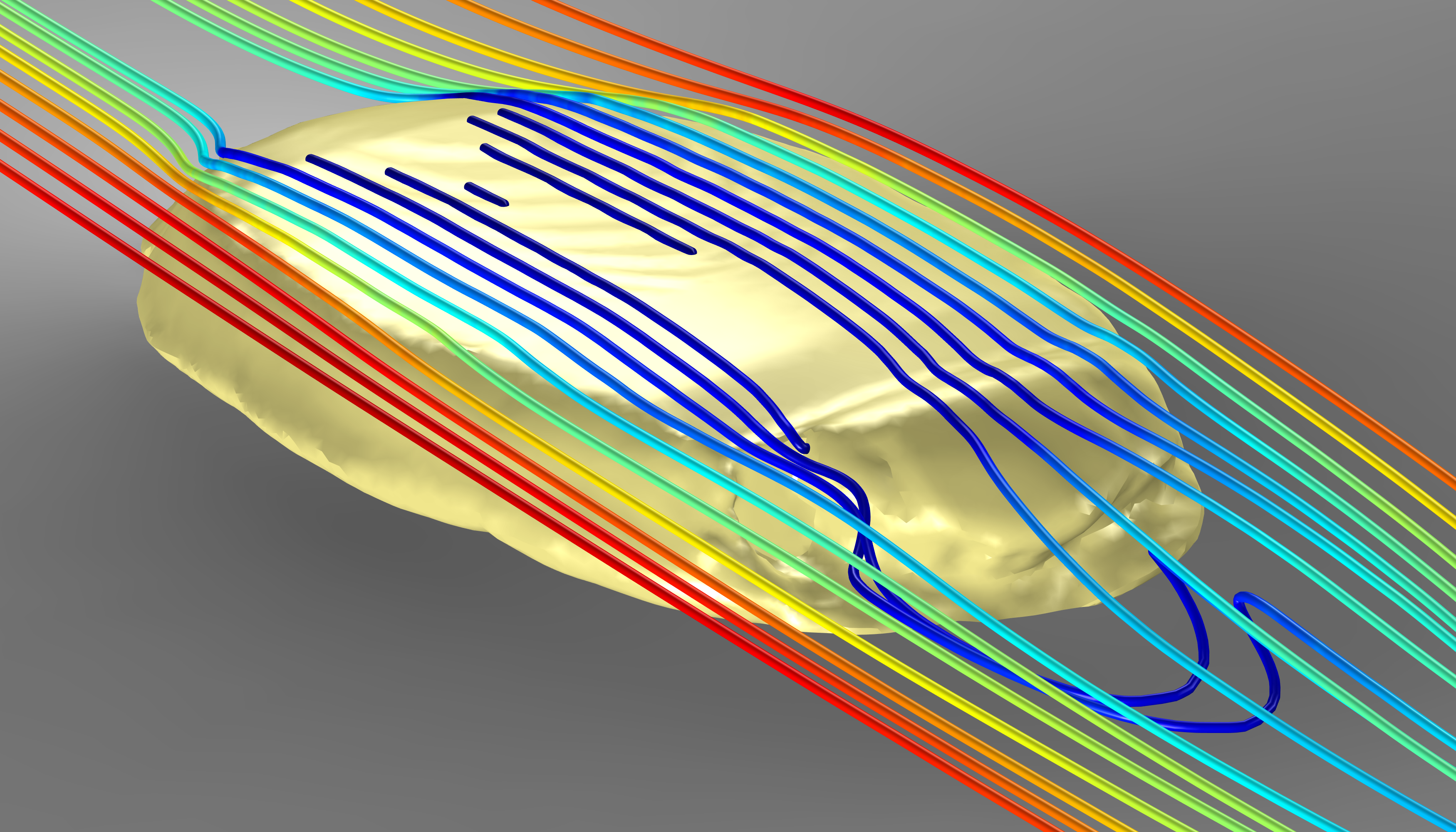
Computer simulation of water flow around a 3-D model of Protocinctus mansillaensis. Image credit – Imran Rahman
The second study, also lead by Dr. Imran Rahman, looked at another primitive echinoderm from the Carboniferous of China (323 million years ago). Using the Swiss Light Source synchrotron technology where x-rays are used to study the internal structure of materials such as fossils. This study looked at an extinct blastoid (a type of echinoderm) in an early-development stage to further understand the internal anatomy of these animals and their evolution. By reconstructing the fossil in 3D, they were able to identify a U-shaped structure which they interpret as the digestive tract. Co-author Dr. Colin Sumrall of the University of Tennessee, Knoxville said: “The results have highlighted a number of previously unknown differences between the fossil and its living relatives. This has forced us to rethink our ideas about how the digestive system evolved in echinoderms.” This study was published in Biology Letters.
Top image: Reconstruction of Protocinctus mansillaensis in life position. Image credit – Oscar Sanisidro.
Related Posts
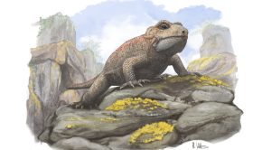
A New Early Reptile With Bizarre Teeth From The Triassic of England →
Episode 161: Notosuchians →
200 Years of Dinosaurs →