Episode 120: Naked Ammonite
It wouldn’t be outlandish to state that many a fossil collection has started with the acquisition of an ammonite. Their planispiral shells (termed a conch) are instantly recognisable and since that conch was originally composed of the relatively hard mineral aragonite, they better lend themselves to the fossilisation process.
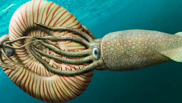
But how much do we actually know about the animal that produces the conch? We might be able to make superficial inferences based on comparisons with the modern Nautilus, but ammonites are actually closer related to squid and octopuses.
So could you recognise an ammonite without its shell?
Prof. Christian Klug of the University of Zurich has recently described just that: a naked ammonite. In this episode, we learn about ammonite soft body anatomy and sink our teeth into the mystery of how this ammonite lost its shell.
Read the original open access paper here: Klug, C., Schweigert, G., Tischlinger, H. et al. Failed prey or peculiar necrolysis? Isolated ammonite soft body from the Late Jurassic of Eichstätt (Germany) with complete digestive tract and male reproductive organs. Swiss J Palaeontol 140, 3 (2021).
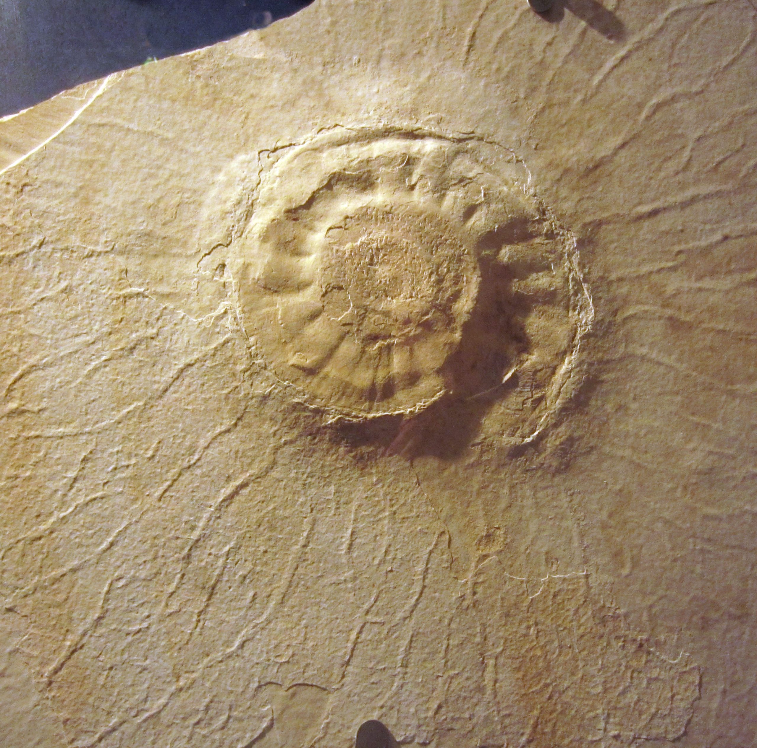
Big perisphinctid ammonite (Late Jurassic) from the ‘Solnhofener Plattenkalke’ in the Museum Solnhofen.
Image: Christian Klug
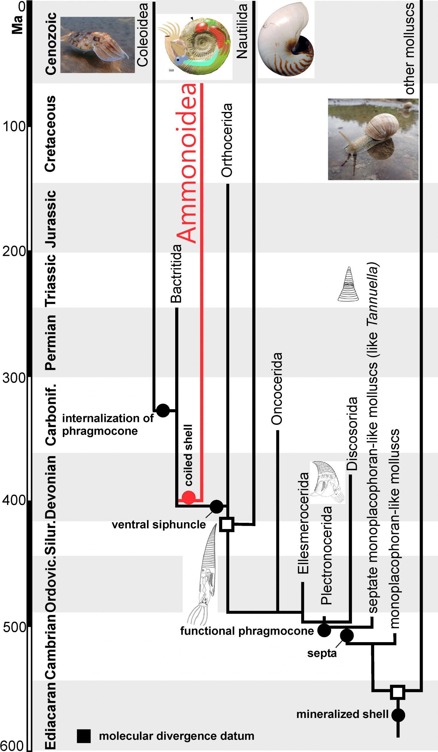
Modified after Kröger et al. (2011: Cephalopod origin and evolution. In Bioessays).
Image: Christian Klug
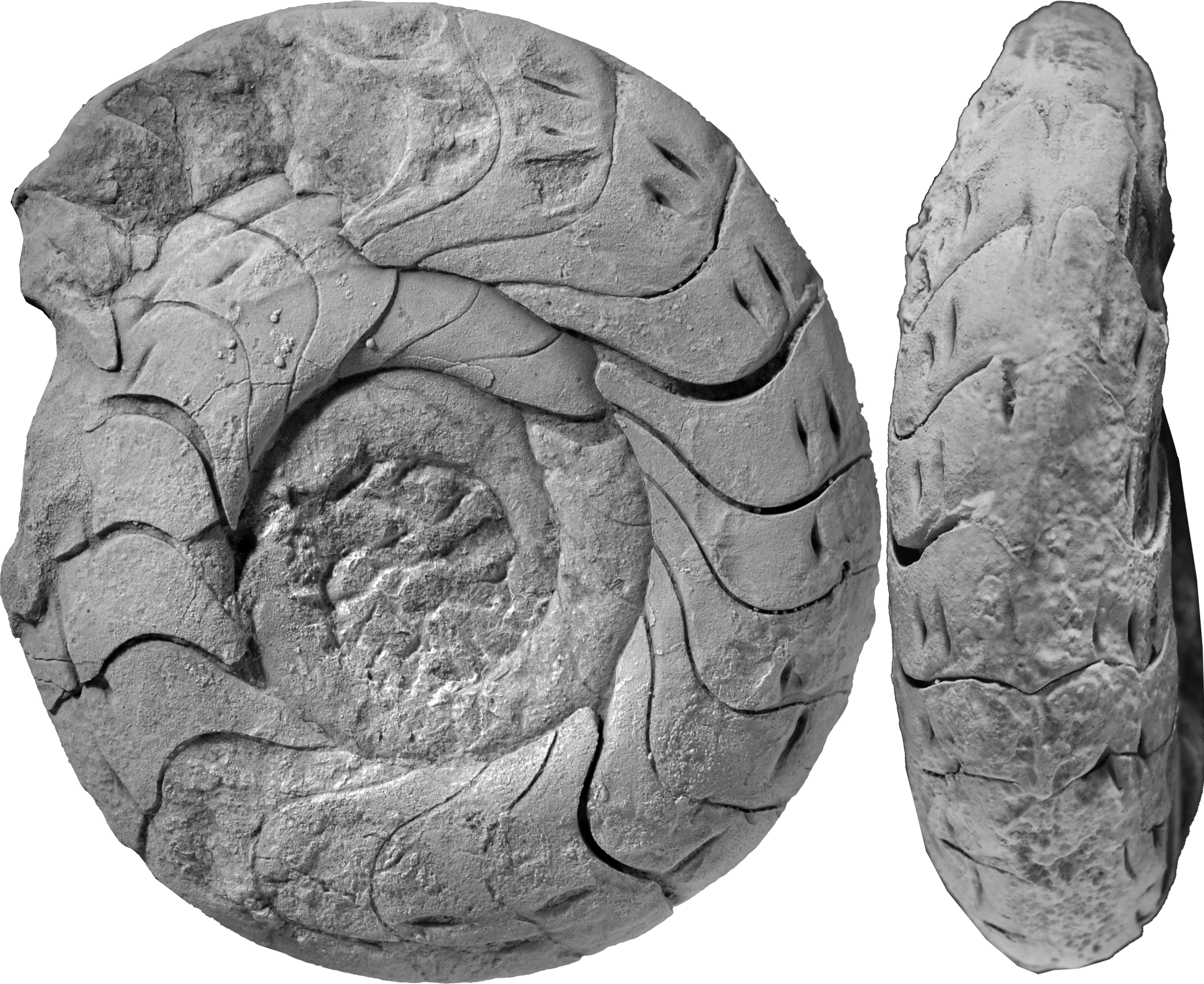
Zigzag-sutures of the Frasnian (Late Devonian) ammonoid Beloceras from the eastern Anti-Atlas of Morocco.
Image: Christian Klug
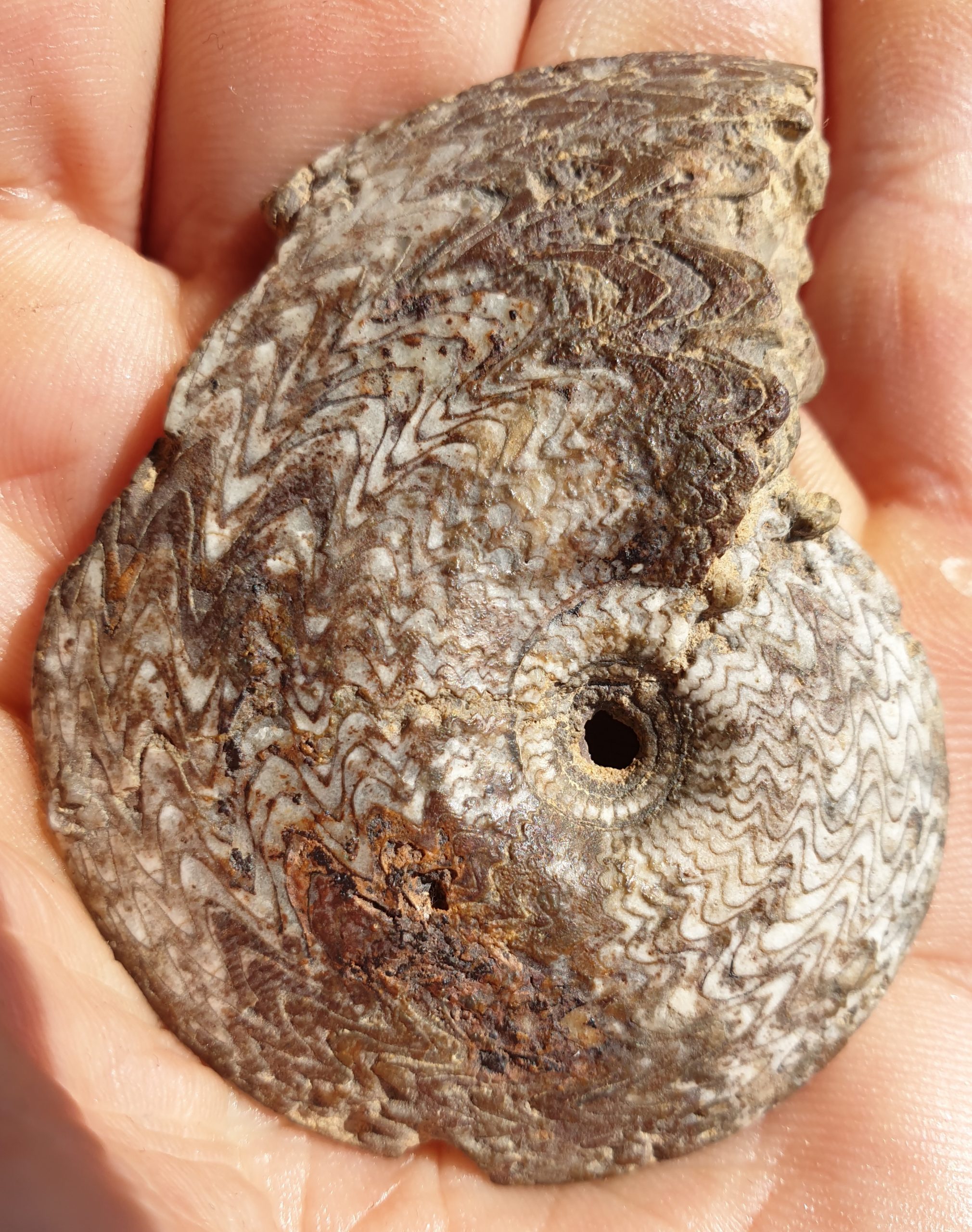
Internal mould of the late Emsian (Early Devonian) ammonoid Sellanarcestes neglectus from the eastern Anti-Atlas (Morocco). Diameter 55 mm.
Image: Christian Klug
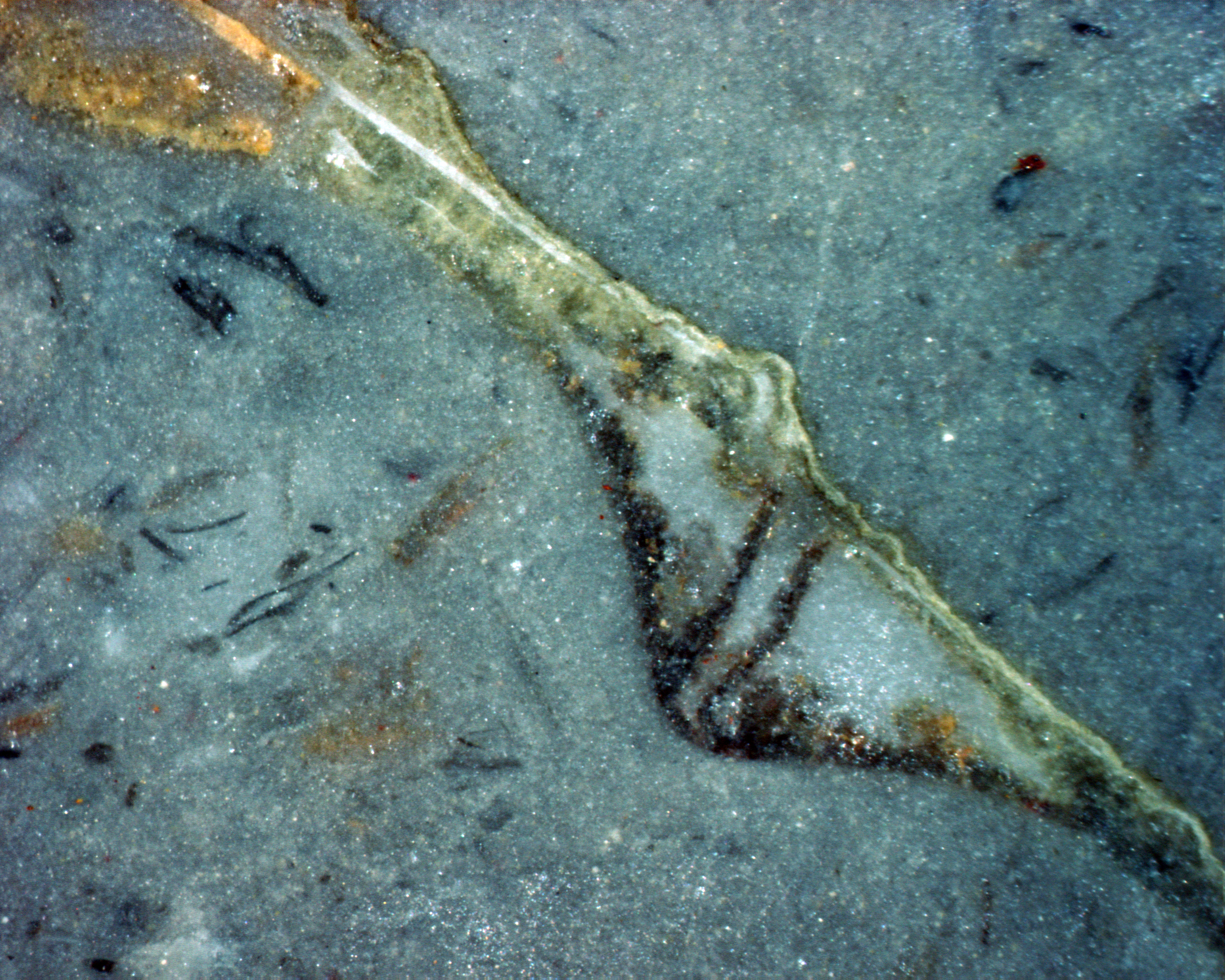
Cross section through a fixed pearl surrounding a small tube (tube about 1 mm long) in the ammonoid Sellanarcestes from the late Emsian (Early Devonian) of Oufrane (Morocco).
Image: Christian Klug
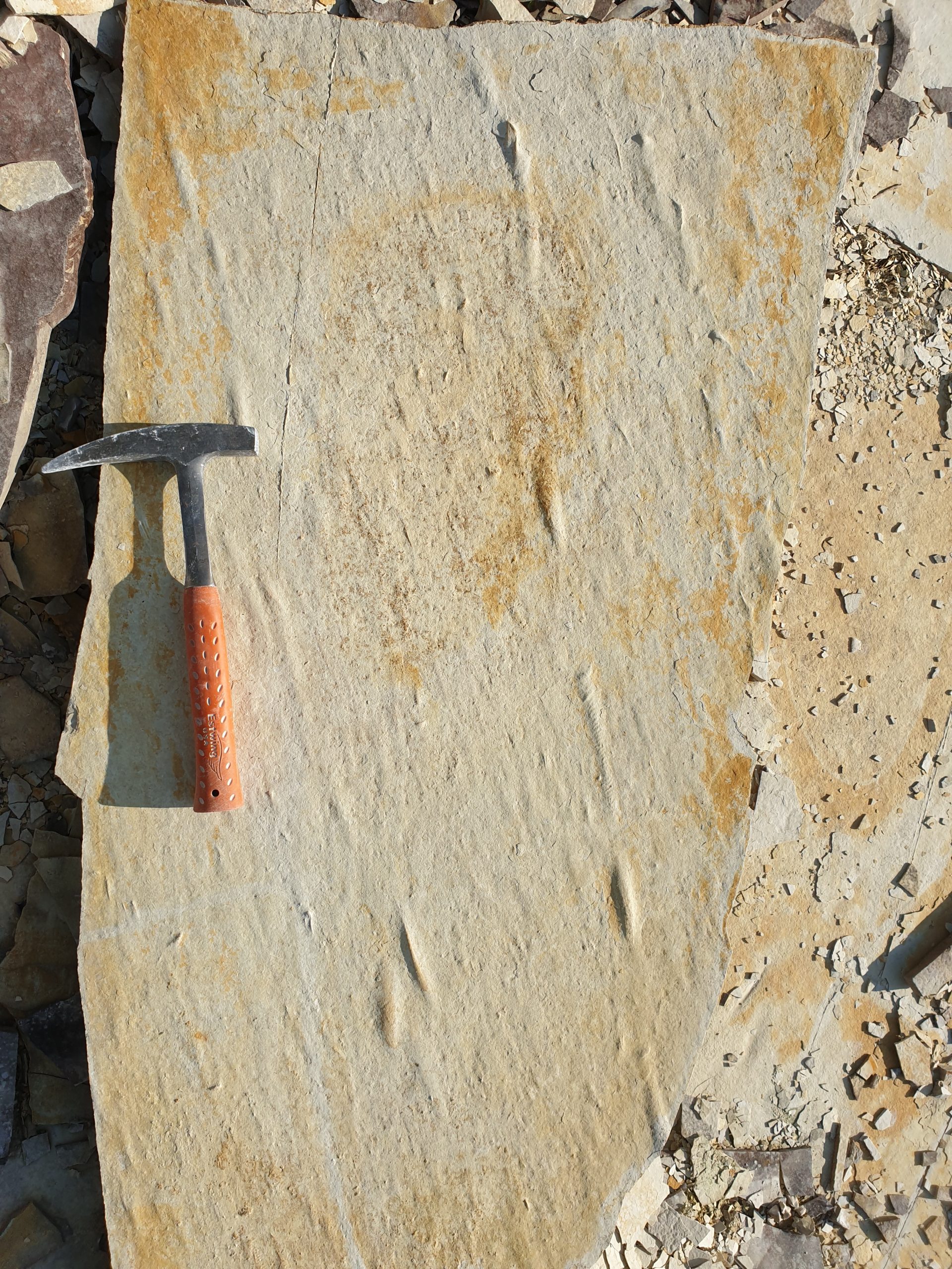
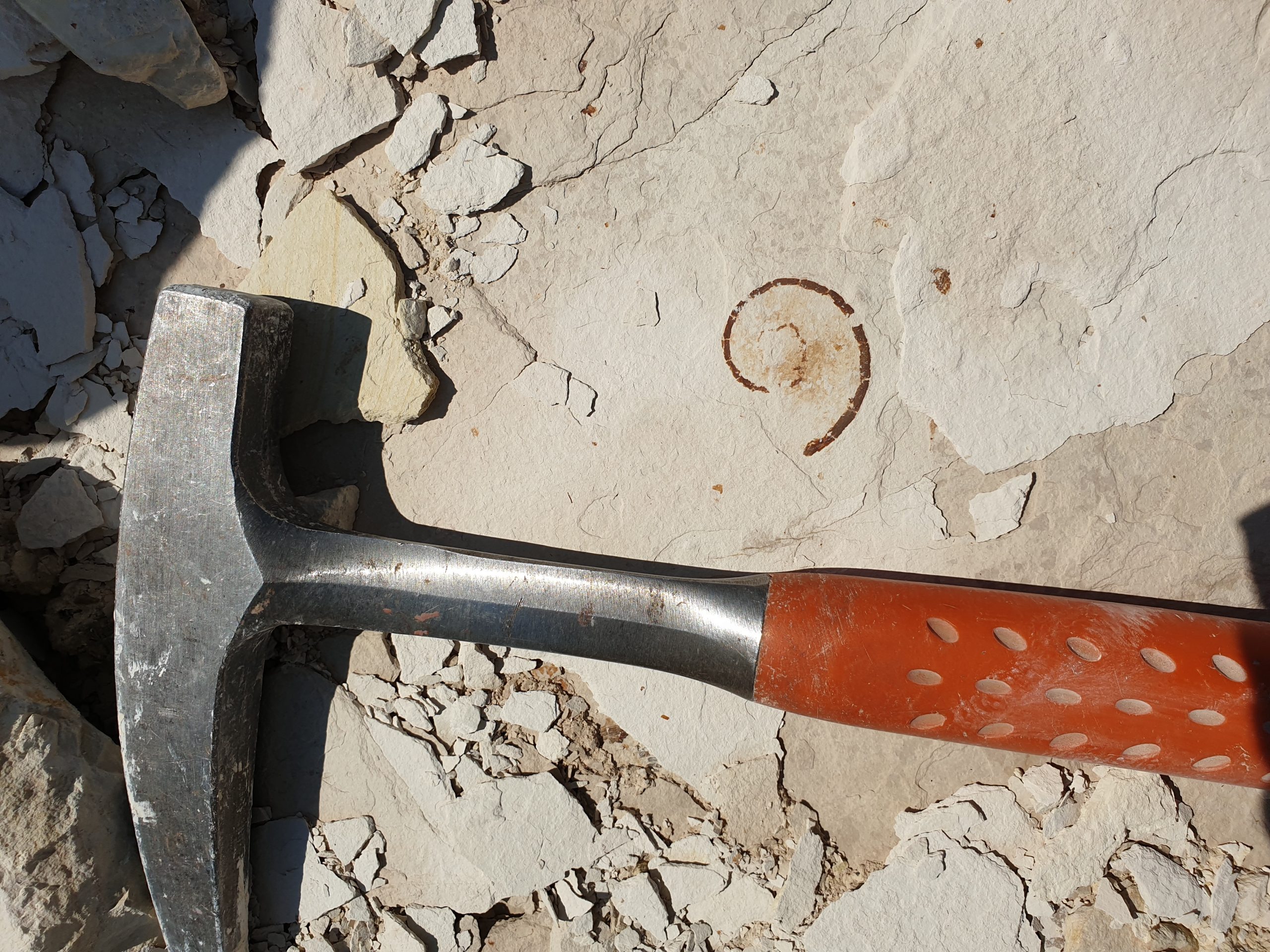
Roll marks of ammonites from the “Solnhofen Plattenkalk” of Painten.
Image: Christian Klug
Jurassic ammonite in the Kimmeridgian Plattenkalk of Painten in siphuncle-preservation (flattened otherwise).
Image: Christian Klug
Image: Christian Klug
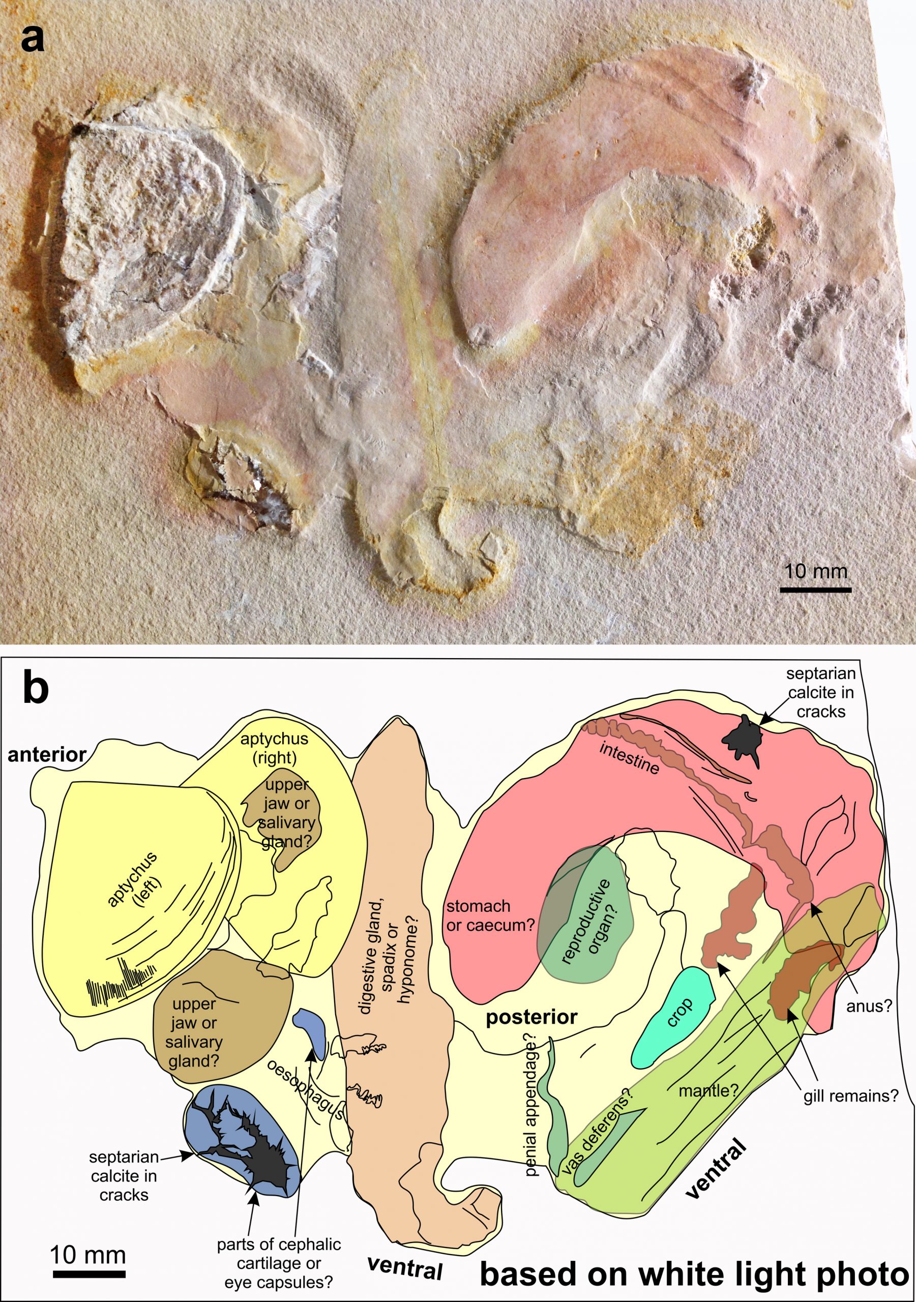
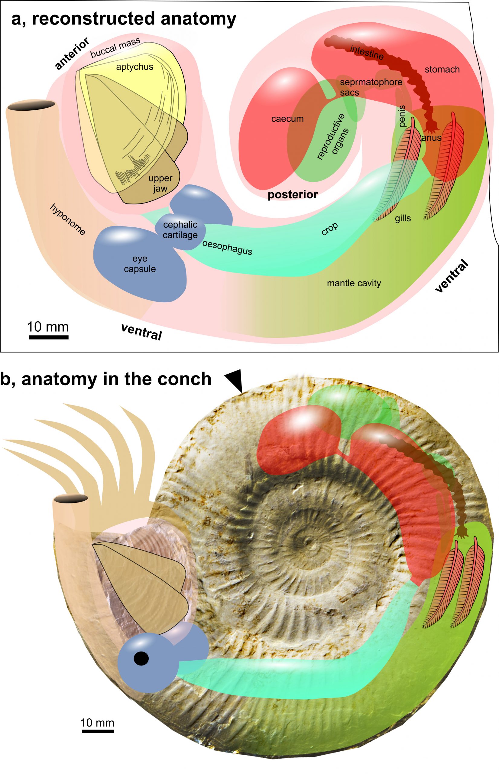
Soft parts of Subplanites sp. with a Strigogranulaptychus sp. from the early Tithonian of Wintershof near Eichstätt (Germany); SMNS 70,610. a Photo taken under white light. b Line drawing of the structures visible in the white light photo (a) with possible interpretations.
Original Illustration from Klug et al. (2021: Fig. 1)
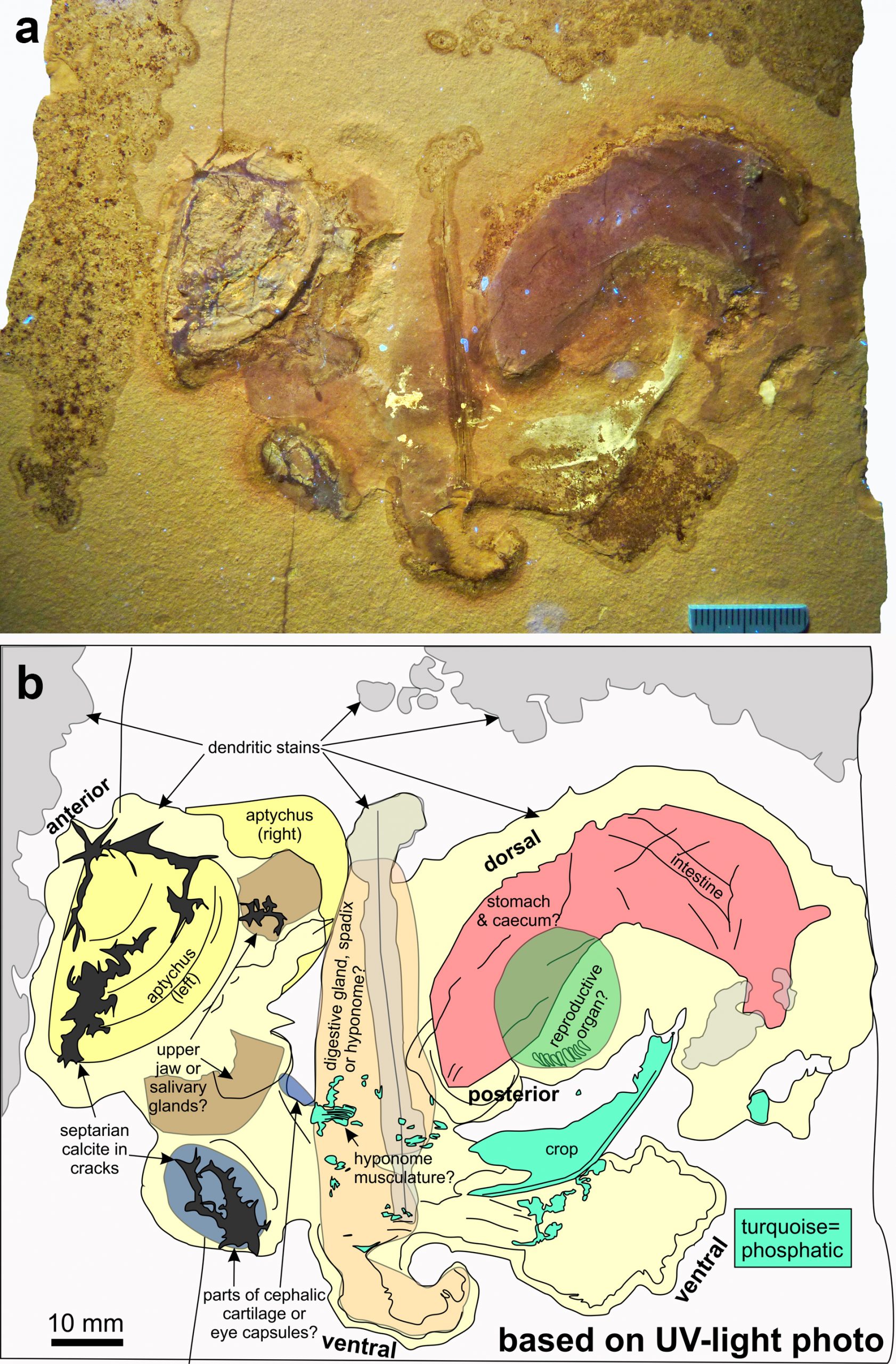
Soft parts of Subplanites sp. with a Strigogranulaptychus sp. from the early Tithonian of Wintershof near Eichstätt (Germany); SMNS 70610. a Photo taken under UV light. b Line drawing of the structures visible in the UV photo (a) with possible interpretations.
Original Illustration from Klug et al. (2021: Fig. 2)
Original Illustration from Klug et al. (2021: Fig. 6)
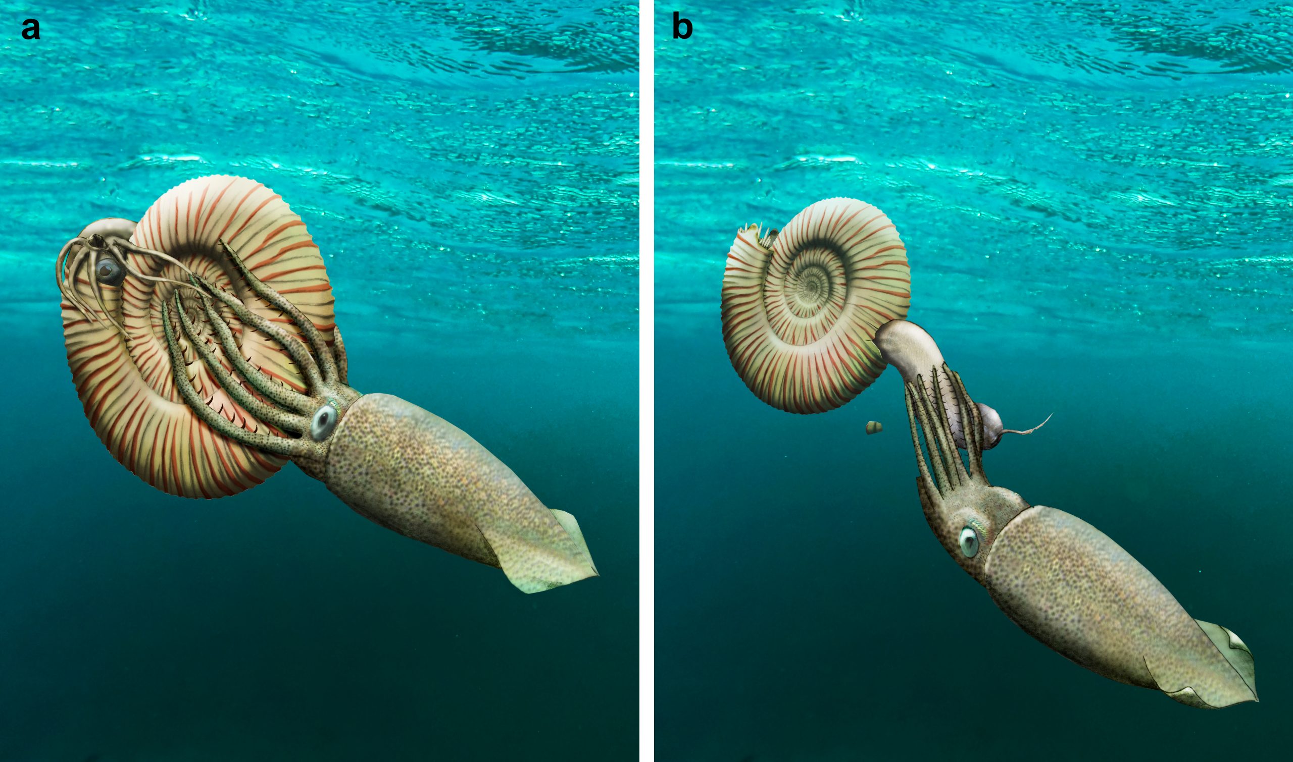
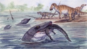
Reconstruction of the possible events that might have led to the extraction of the ammonite soft parts. A coleoid (belemnite or octopus ancestor) bit a hole in the rear of the body chamber and pulled out the soft parts; subsequently, it might have dropped them.
Original Illustration from Klug et al. (2021: Fig. 7)
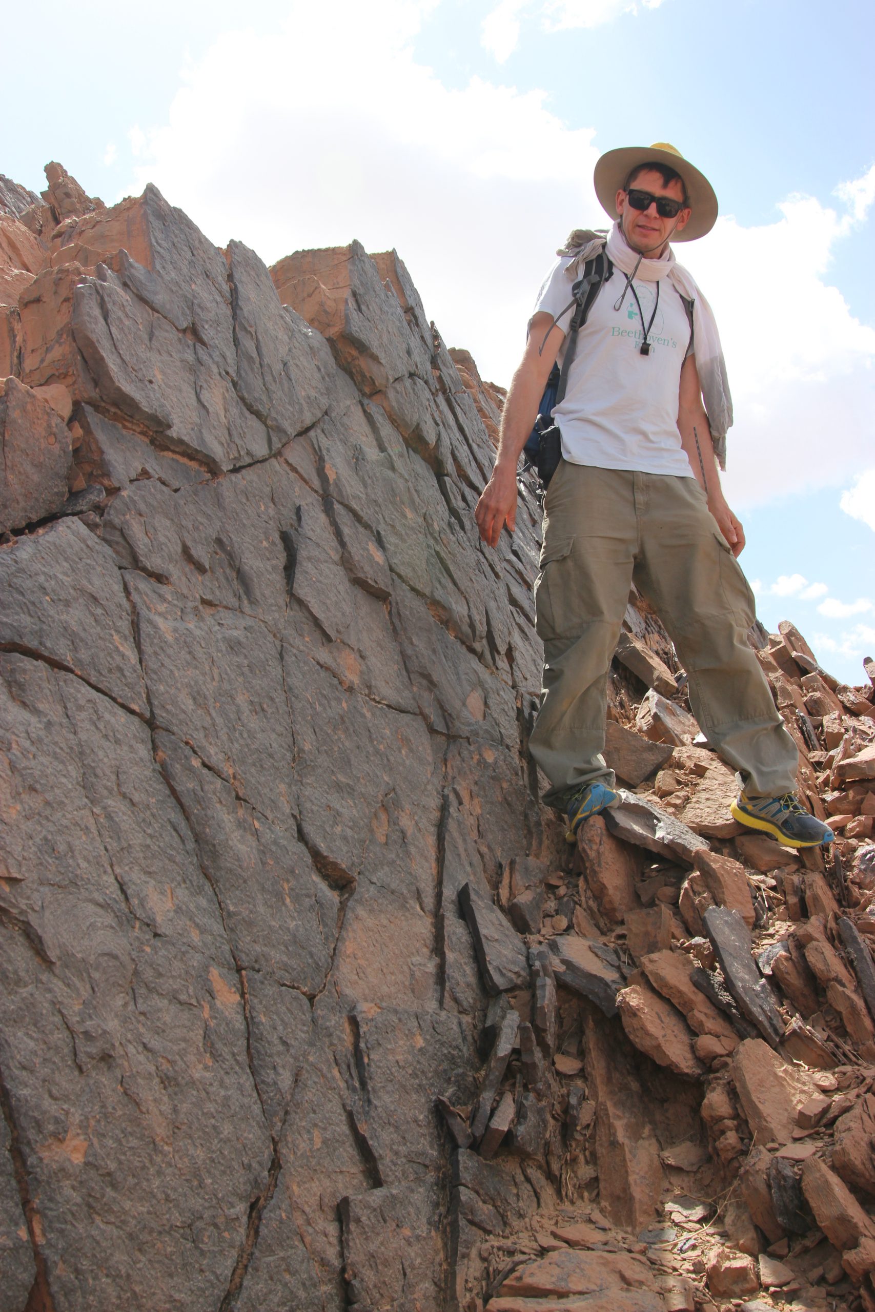
Image: Laura Heck
Related Posts
Episode 174: A History of Dinosaurs in 50 Fossils →
Episode 172: Rhynchocephalians →
Episode 171: Freshwater Mosasaurs →