Episode 89: Teeth and herbivory in reptiles
Tooth shape and arrangement is strongly linked with diet, and palaeontologists often use teeth to determine what kind of food an animal may have been eating. Carnivorous teeth are generally more simple, while herbivorous teeth are more complicated. We know that herbivory evolved later, but how did the dentition of herbivores evolve? What kind of variation exists in herbivorous dentition?
In this episode, we speak with Dr Aaron LeBlanc, a Killam Postdoctoral Fellow at the University of Alberta. His research focuses on the evolution and development of teeth in amniotes, including some of his PhD work on the development of the dental system in herbivores, which we discuss in detail here, as well as the evolution of the mammalian system, which earned him the Alfred S. Romer Student Prize at last year’s SVP in Calgary.
Podcast: Download (Duration: 56:51 — 79.5MB)
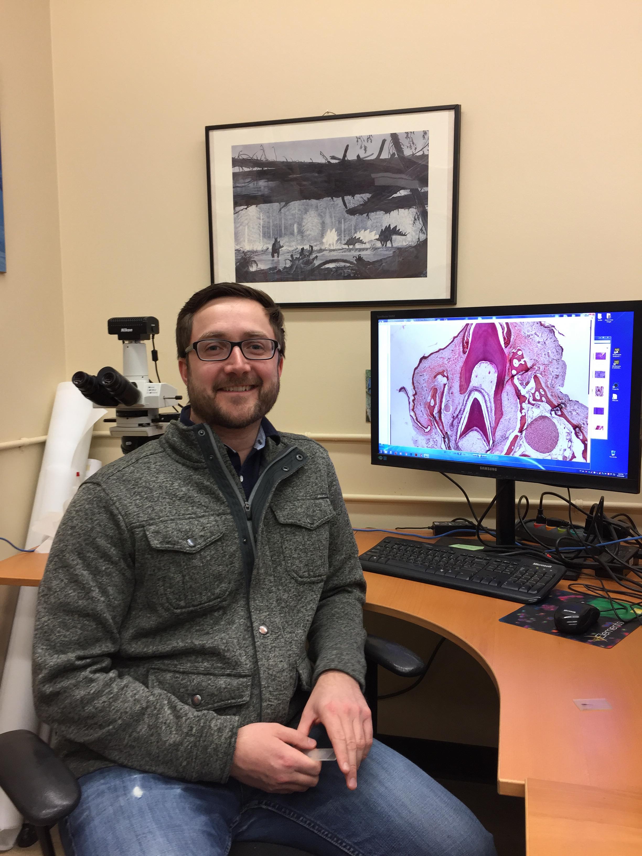
Dr. Aaron LeBlanc with a histological section through the jaw of a modern caiman, showing a functional tooth and a developing replacement tooth
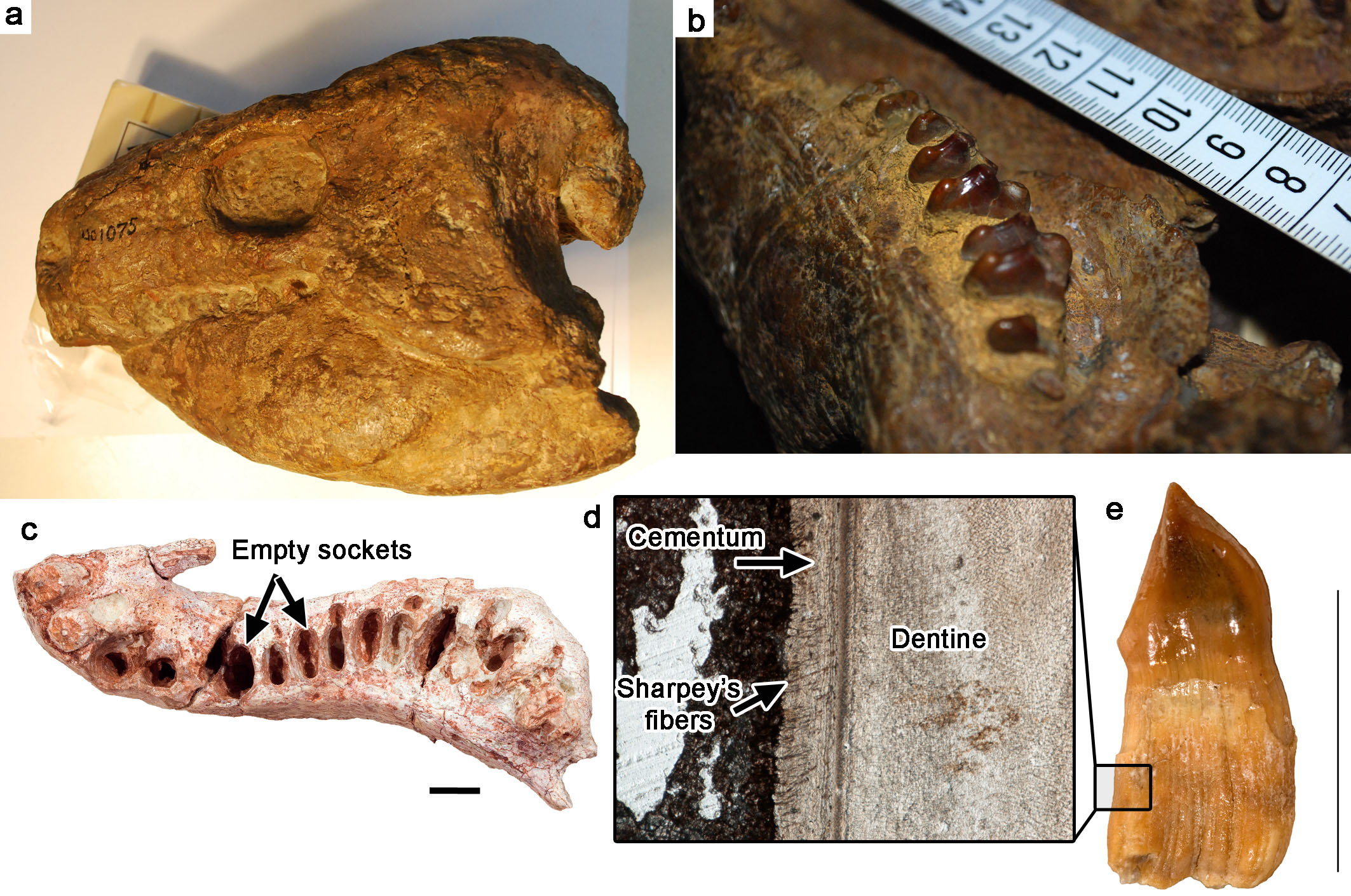
The teeth of the herbivorous tetrapod, Diadectes, one of the earliest tetrapod herbivores from the Late Palaeozoic. a, a skull of Diadectes from the Field Museum in Chicago, U.S.A. b, a close-up of the molariform teeth of a diadectid. c, an upper jaw (maxilla and premaxilla) of a diadectid with empty tooth sockets, showing that soft tissue held the teeth in place in life. d, a thin section of a diadectid tooth, showing the tooth attachment tissues (cementum) and the anchoring points (Sharpey’s fibers) along the root surface for a ligament that would have held the tooth in place. e, an isolated, worn diadectid tooth.
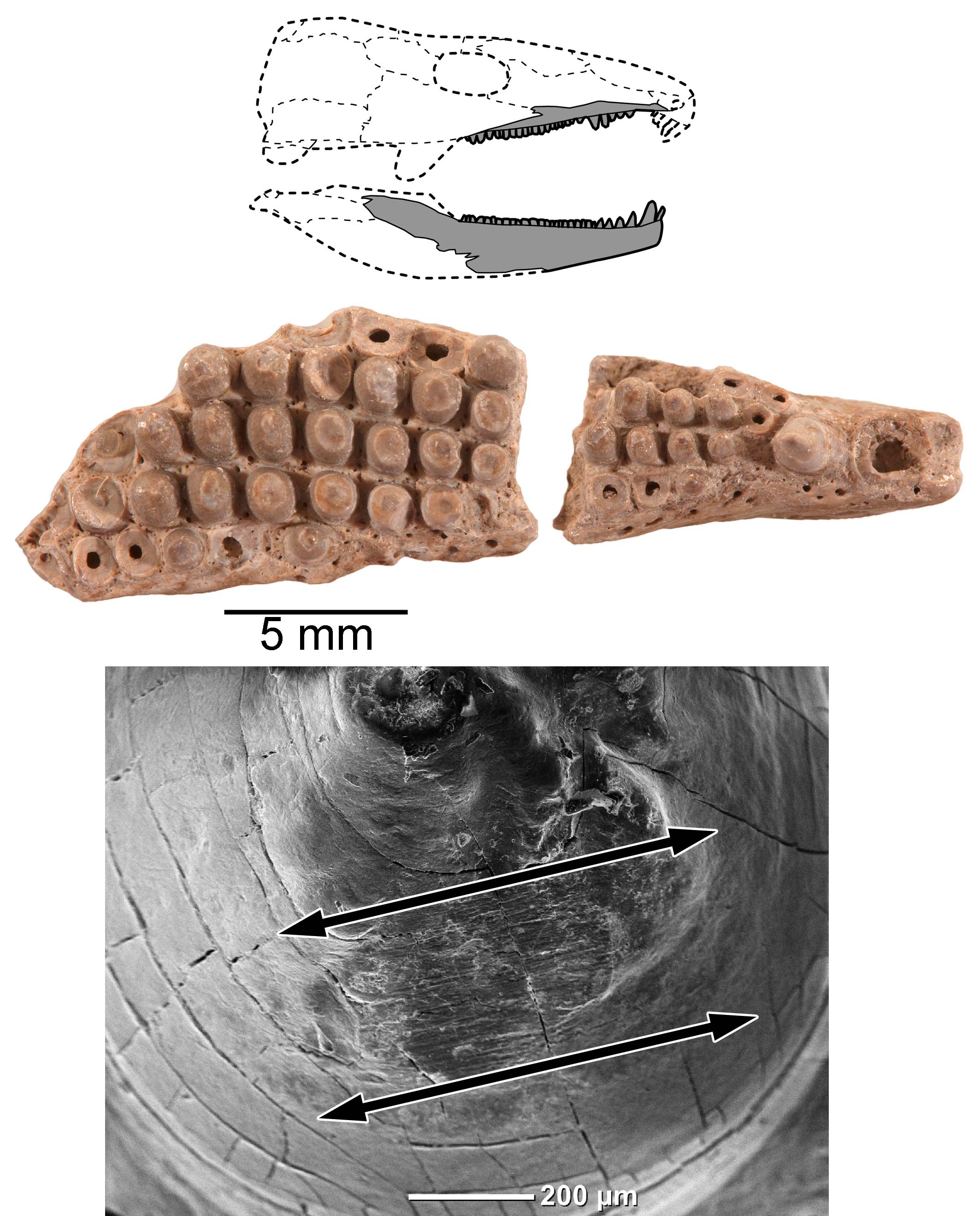
Tooth wear in the multiple tooth-rowed captorhinid reptile Captorhinikos from Oklahoma U.S.A. Striations along the worn teeth in Scanning Electron Microscope images (bottom) show that the lower teeth moved forward and backwards across the upper teeth to pulverize tough plant material.
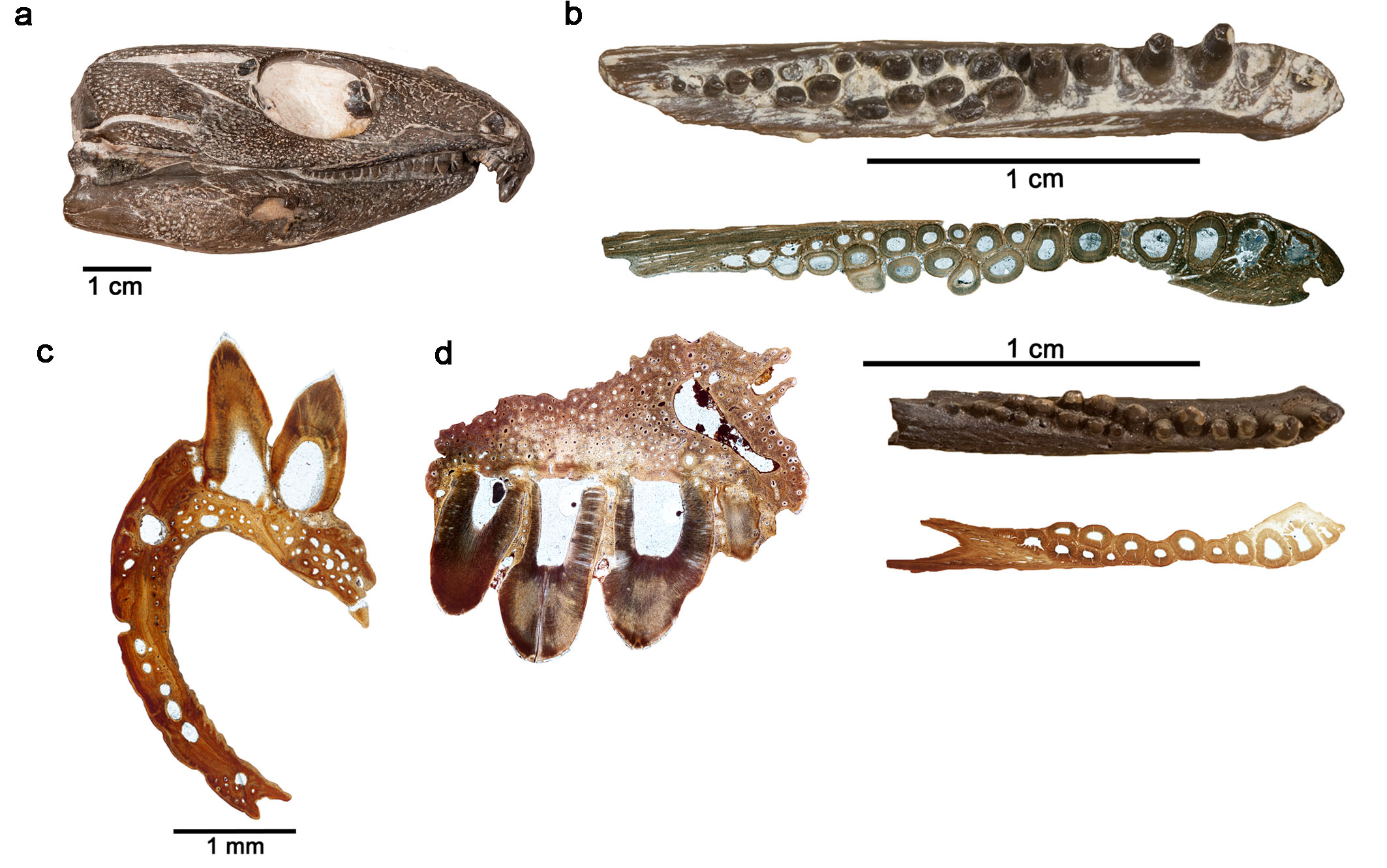
Histological sections of the teeth of the multiple-rowed captorhinid Captorhinus aguti. a, a Captorhinus skull from the Dolese Brothers Quarry in Oklahoma, U.S.A. b, images and thin sections of small and larger C. aguti with multiple tooth rows. c, thin section of the multiple-rowed region of the dentary of C. aguti. d, thin section of the multiple-rowed region of the maxilla. Photos of specimens courtesy of Diane Scott.
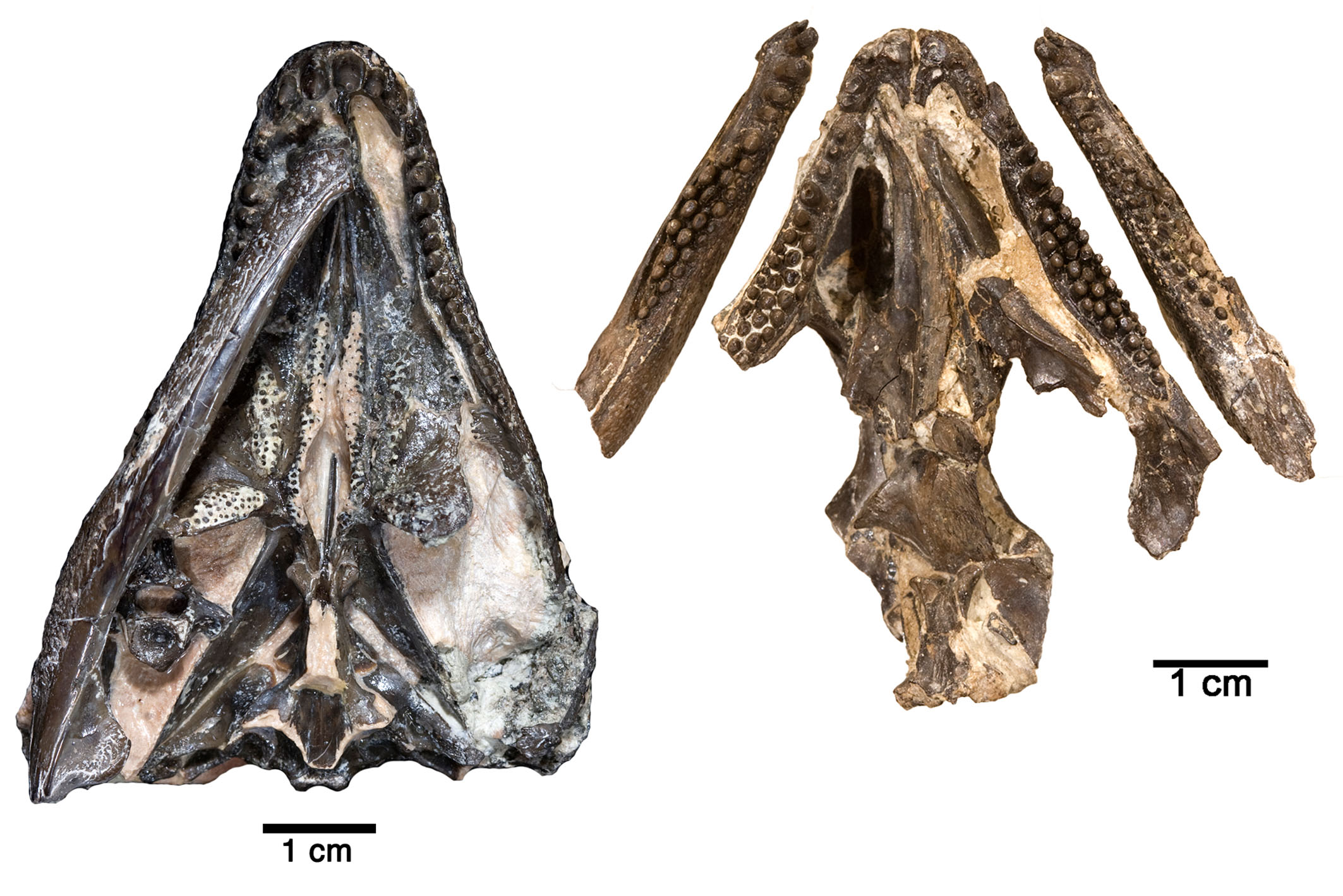
Captorhinus skulls from the Dolese Brothers Quarry in Oklahoma, U.S.A. showing the single (left) and multiple-rowed (right) conditions.
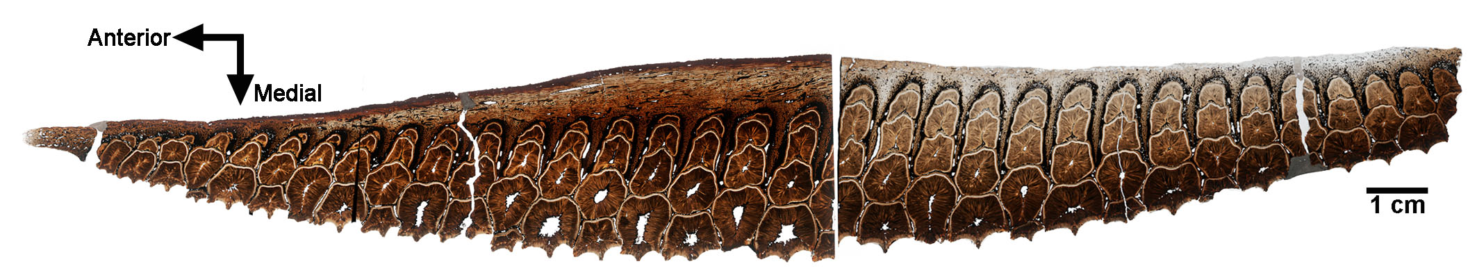
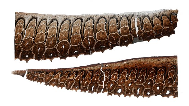
Thin sections across an entire dental battery in the dentary of a hadrosaurid (“duck-billed”) dinosaur. Each hadrosaurid dental battery can consist of hundreds of teeth.
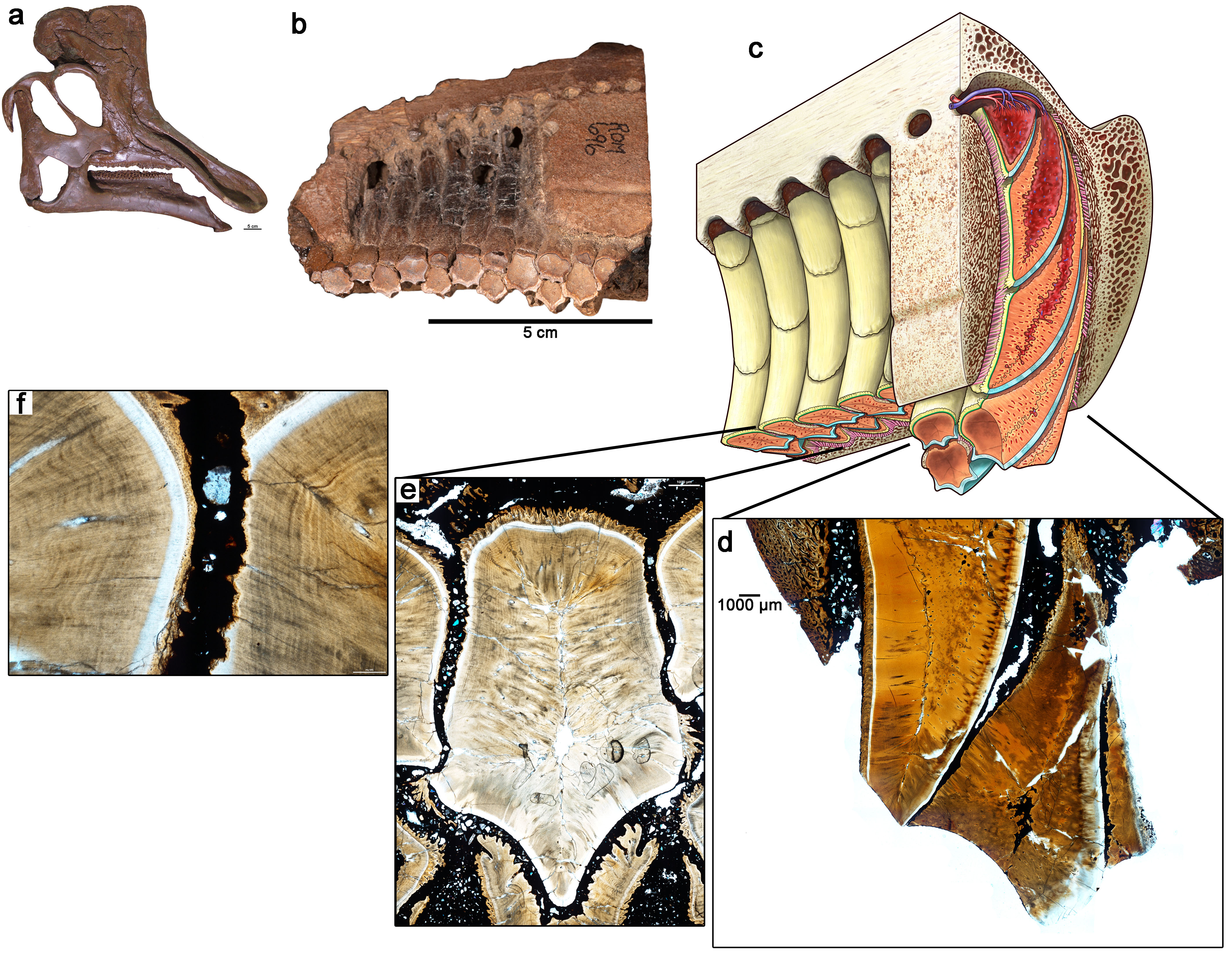
The internal anatomy of a hadrosaurid dental battery as revealed by histology. a, a skull of the hadrosaurid Corythosaurus. b, a lingual view of a hadrosaurid maxillary dental battery, showing the arrangements of the teeth along the grinding surface. c, artist’s reconstruction of the maxillary dental battery (illustration by Danielle Dufault). d, close-up of a thin section through the grinding teeth of the dental battery. Each tooth consists of a solid block of dentine by the time it reaches the grinding surface and can then be ground down completely. e, close-up of a section through a tooth within an intact hadrosaurid dental battery. Note that none of the teeth in the image are in contact with each other, implying that soft tissue would have filled the spaces in between all of the teeth. f, closeup of e showing the sediment-filled space in between the two teeth. This space would have been filled by a ligament in life.
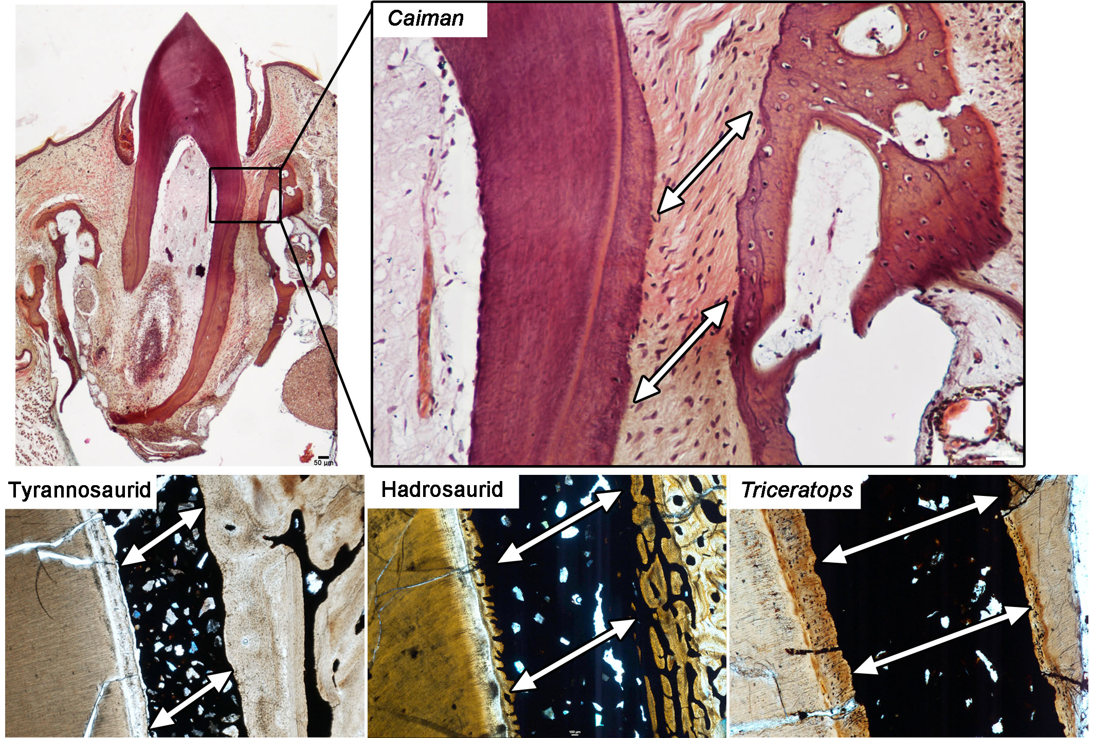
As the only living toothed archosaurs, crocodilians provide the best window into how teeth developed and were anchored into the jaws in extinct dinosaurs. A thin section through the dentary of the extant Caiman sclerops (upper images) shows the ligament that spans the gap between the tooth root and the socket (white arrows), suspending each tooth within the jaws and providing cushioning during a bite. This appears to be the case for dinosaurs as well, because the tooth roots are always separated from the surrounding socket bone, and even neighbouring teeth, by sediment or mineral-filled spaces. Ligaments would have spanned these gaps in life and they would have served similar functions. Among living tetrapods, this ligamentous mode of tooth attachment is also present in mammals.
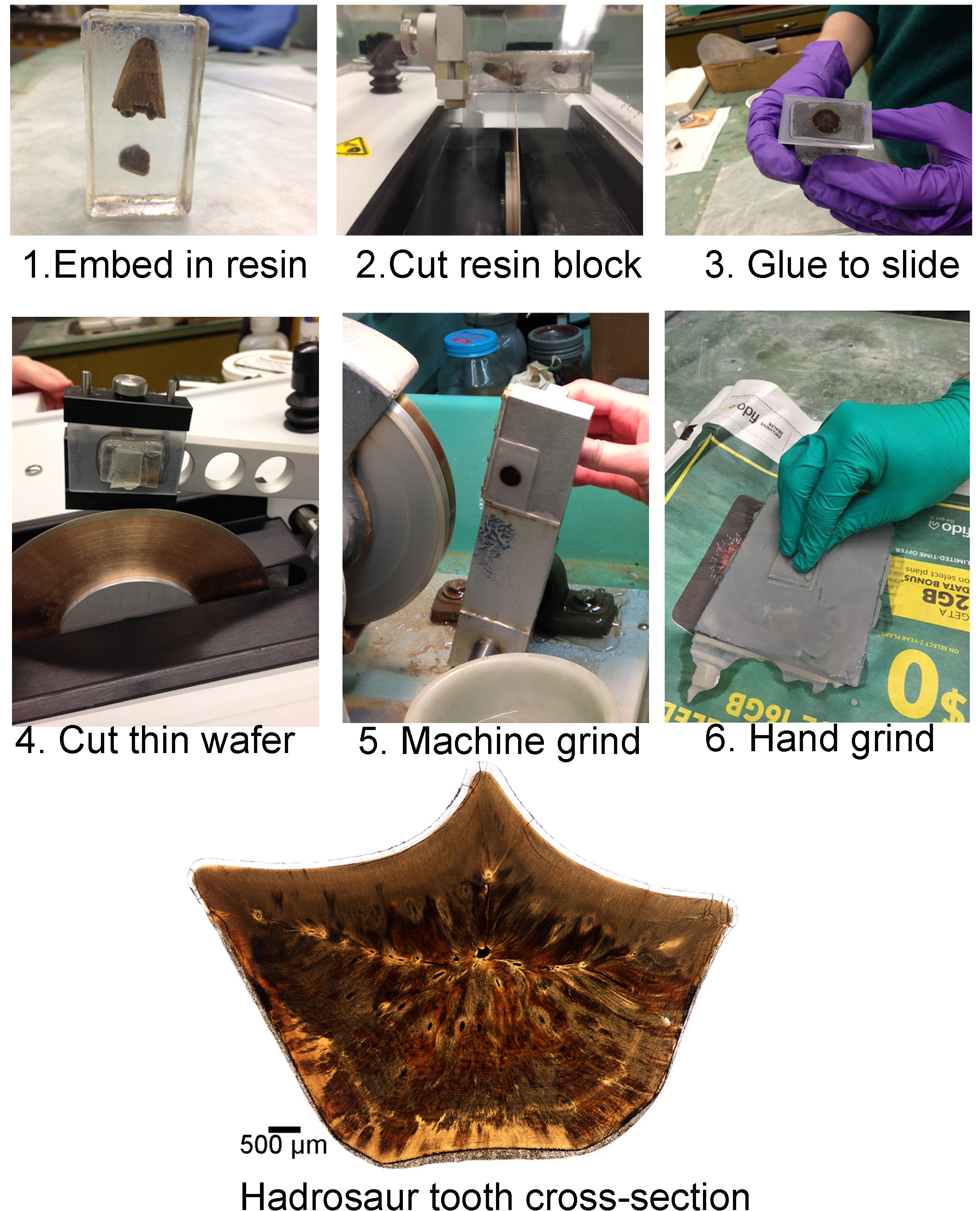
Thin-sectioning procedure.
Related Posts
Episode 160: An introduction to Evolutionary Biology →

Why did Sauropods grow so big? →

Episode 159: An Introduction to Palaeontology →