Episode 15: Micropalaeontology
Perhaps one of the most overlooked areas of palaeontology, within the public eye, is micropalaeontology. Micropalaeontology is an umbrella discipline, covering a diverse range of organisms, with representatives from many of the highest level biological groupings. Although small in size, microfossils prove invaluable for research into palaeoclimatology and are also one of the most commercially applicable groups of fossils.
In this interview we speak to Dr. Giles Miller, Senior Curator of Micropalaeontology at the Natural History Museum (NHM). As each individual group of microfossils could warrant an entire series, this episode serves as an introduction to micropalaeontology. We discuss what it is and some of its applications, all within the context of how the micropalaeontology collection at the NHM is used.
You can keep up to date on the research activities of the micropalaeontology department by following the Curator of Micropalaeontology’s blog, where you can also find further information about case studies covered in this episode.
Podcast: Download (Duration: 49:08 — 67.5MB)
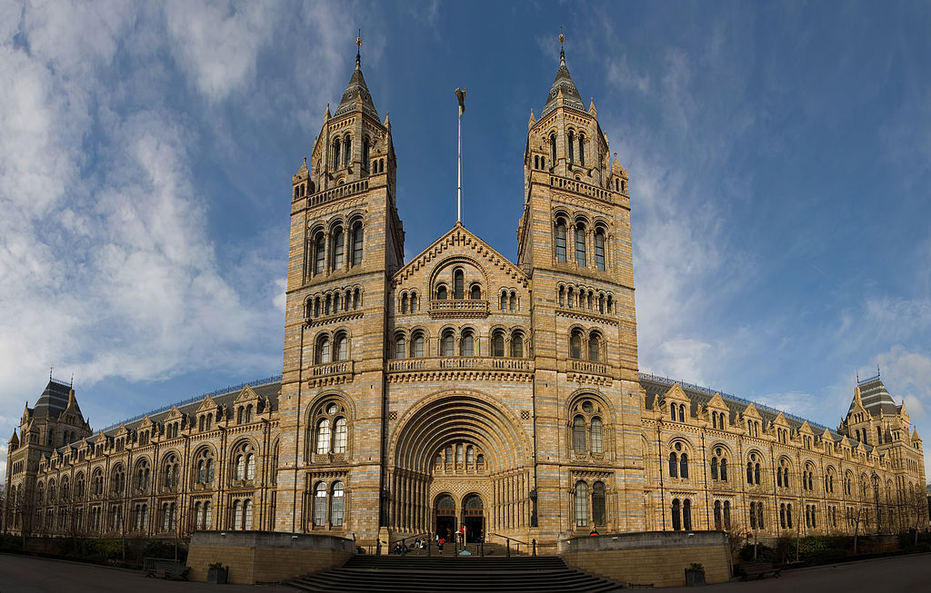
Many museums, such as the Natural History Museum, London, UK, not only have public displays of artefacts, but also possess large collections ‘behind the scenes’ for conservation and research. Image credit: Photo by DAVID ILIFF. License CCBYSA 3.0
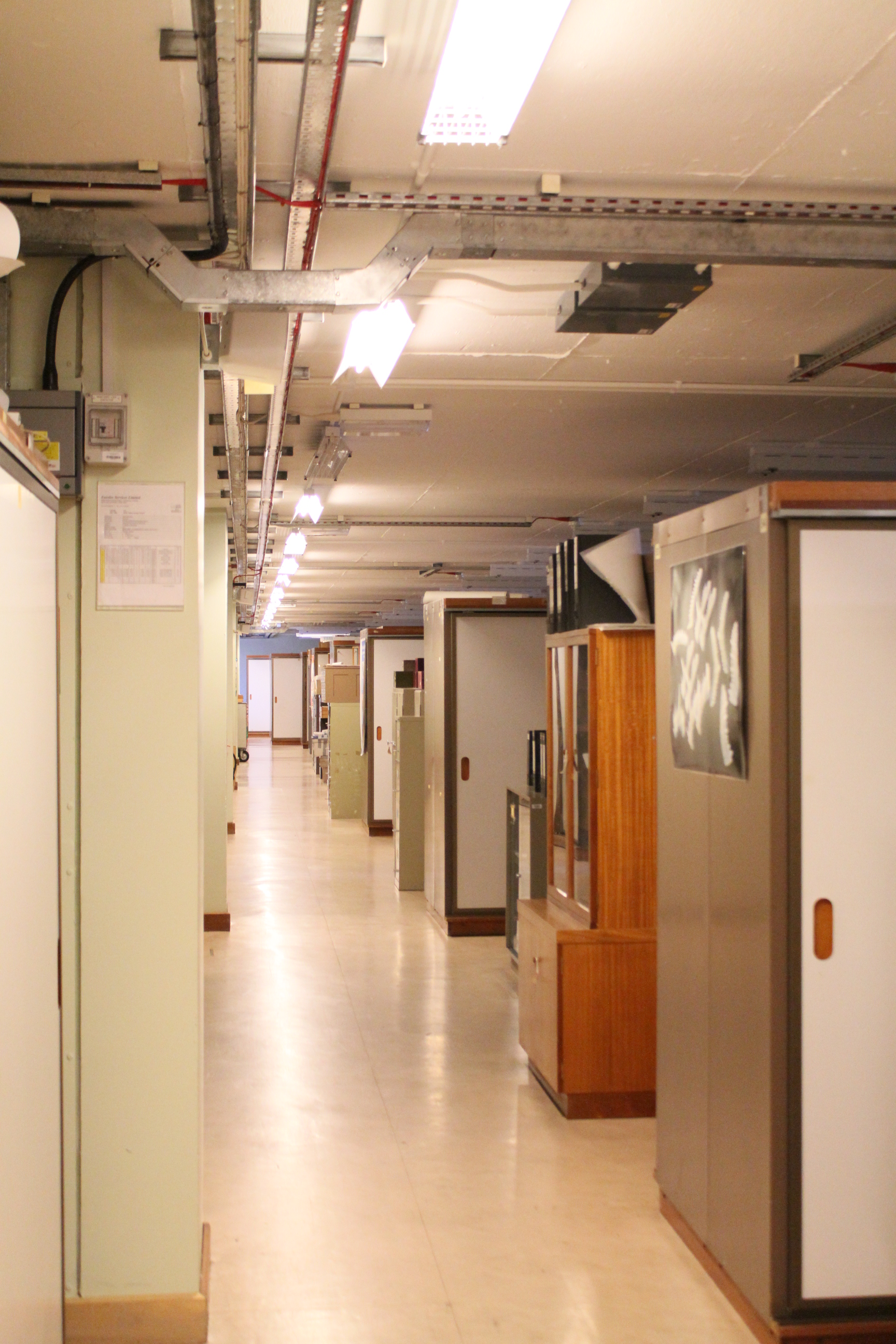
The NHM holds one of, if not the, largest and most important collections of fossils in the UK. As part of this collection, the NHM possesses a large microfossil collection. Image copyright NHM and reproduced with permission.
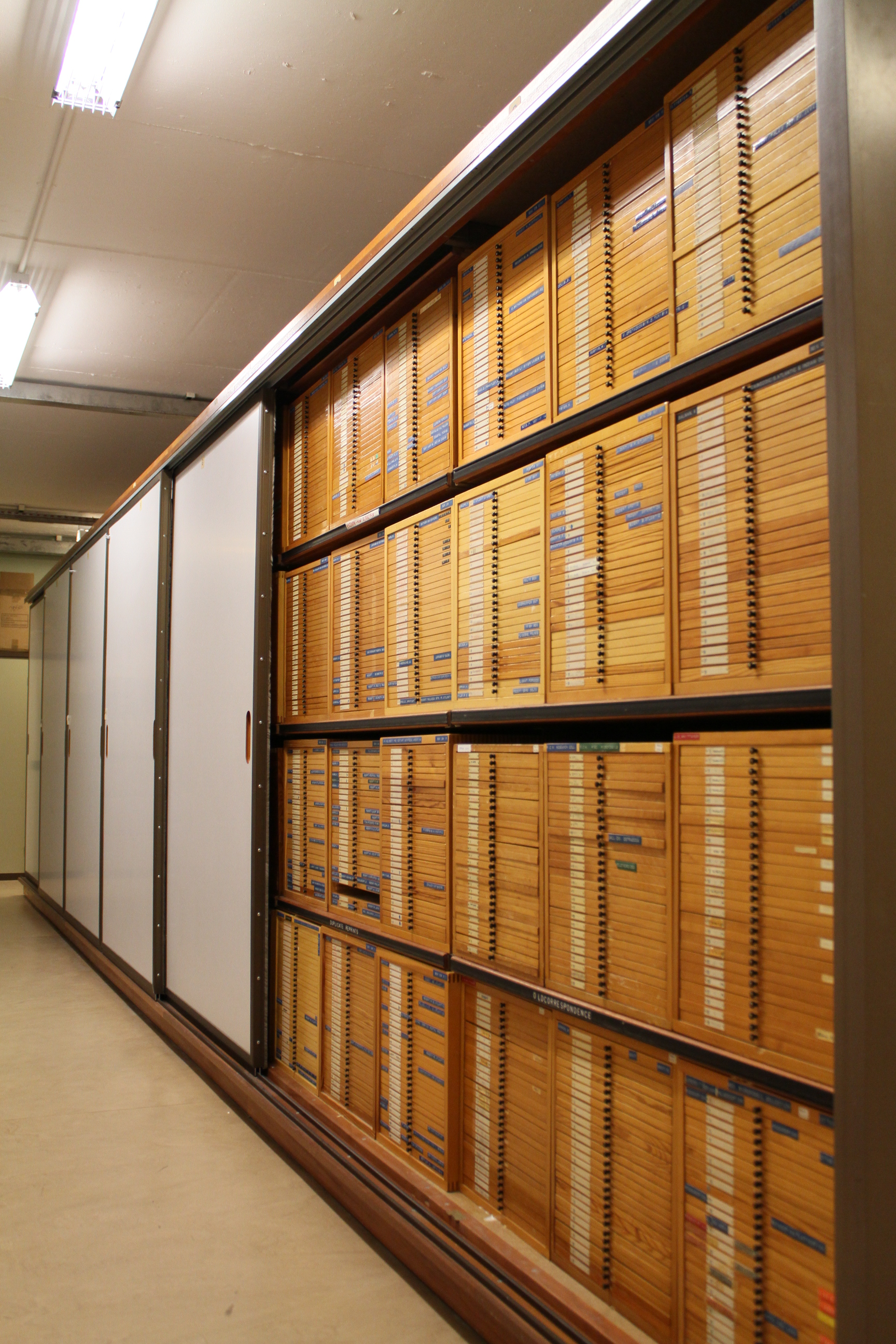
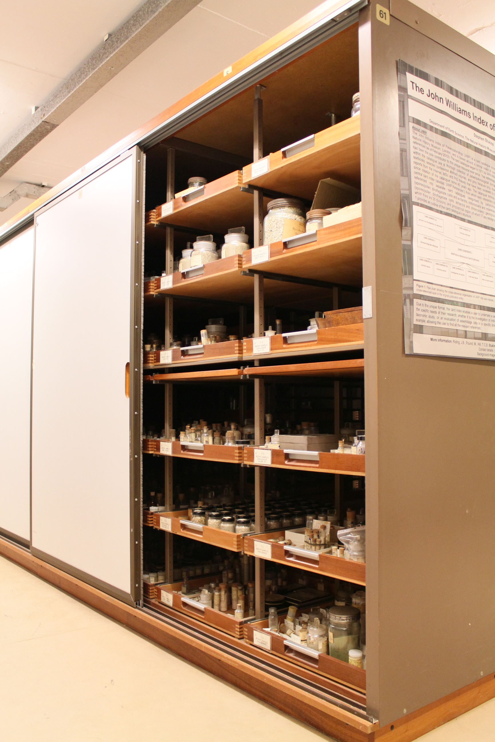
The collection is typically housed in draws where they are made available for researchers. Image copyright NHM and reproduced with permission.
Each drawer can contain many slides. These may have been arranged into taxonomic or geographic groups. Image copyright NHM and reproduced with permission.
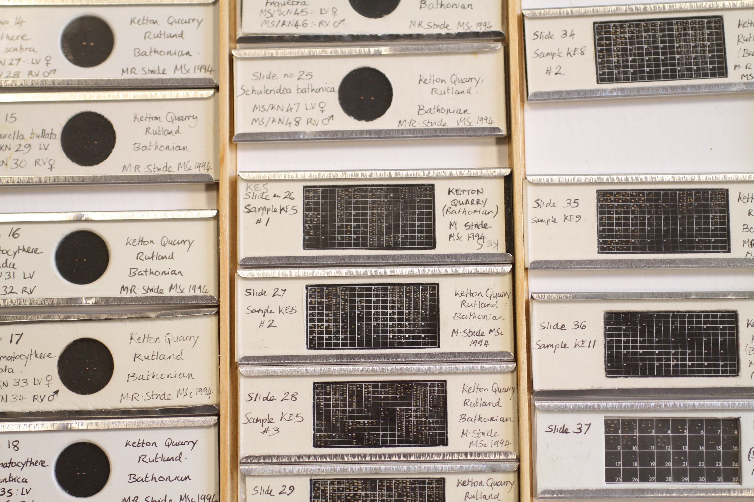
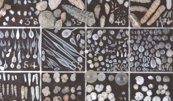
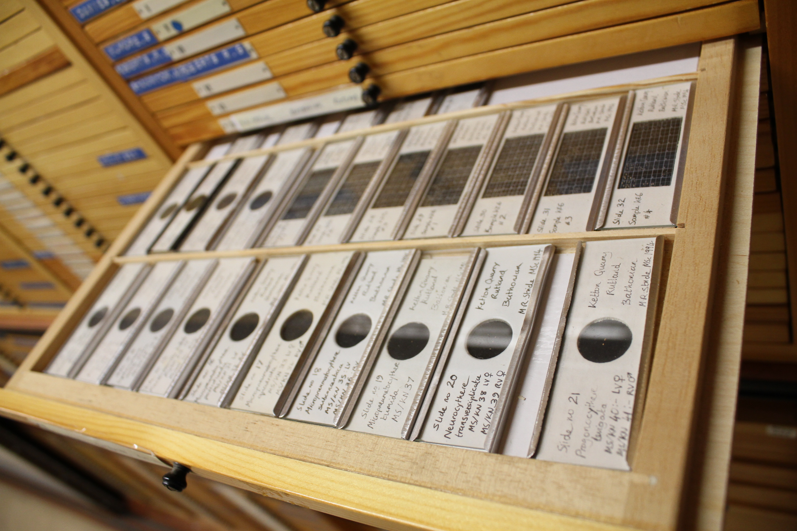
Each slide can contain hundreds of specimens. Some of these are ‘type specimens’, meaning that they are the morphological standard for that particular species. These fossils are used primarily for reference when identifying new and existing species. Image copyright NHM and reproduced with permission.
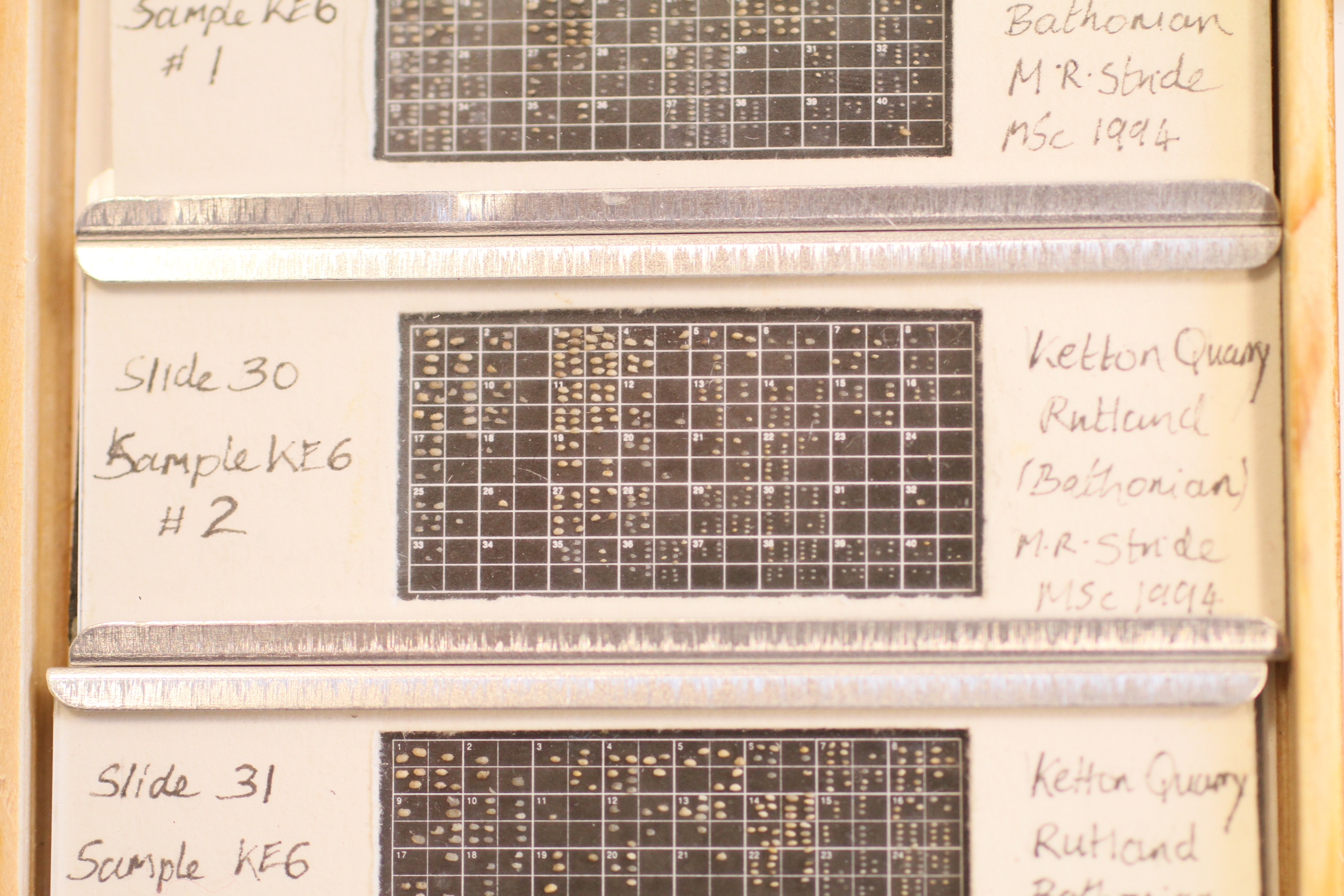
Grid slides, such as this, help micropalaeontologists quickly and easily sort specimens. Each fossil is glued onto the slide using various glues. This slide contains ostracods from the Middle Jurassic of the UK. Image copyright NHM and reproduced with permission.
Microfossils are also stored as ‘residues’. These are somewhat-refined geological samples that preserve relative abundances of microfossils. These are useful for determining palaeoecologies, which in turn are used in palaeoenvironmental reconstructions. Image copyright NHM and reproduced with permission.
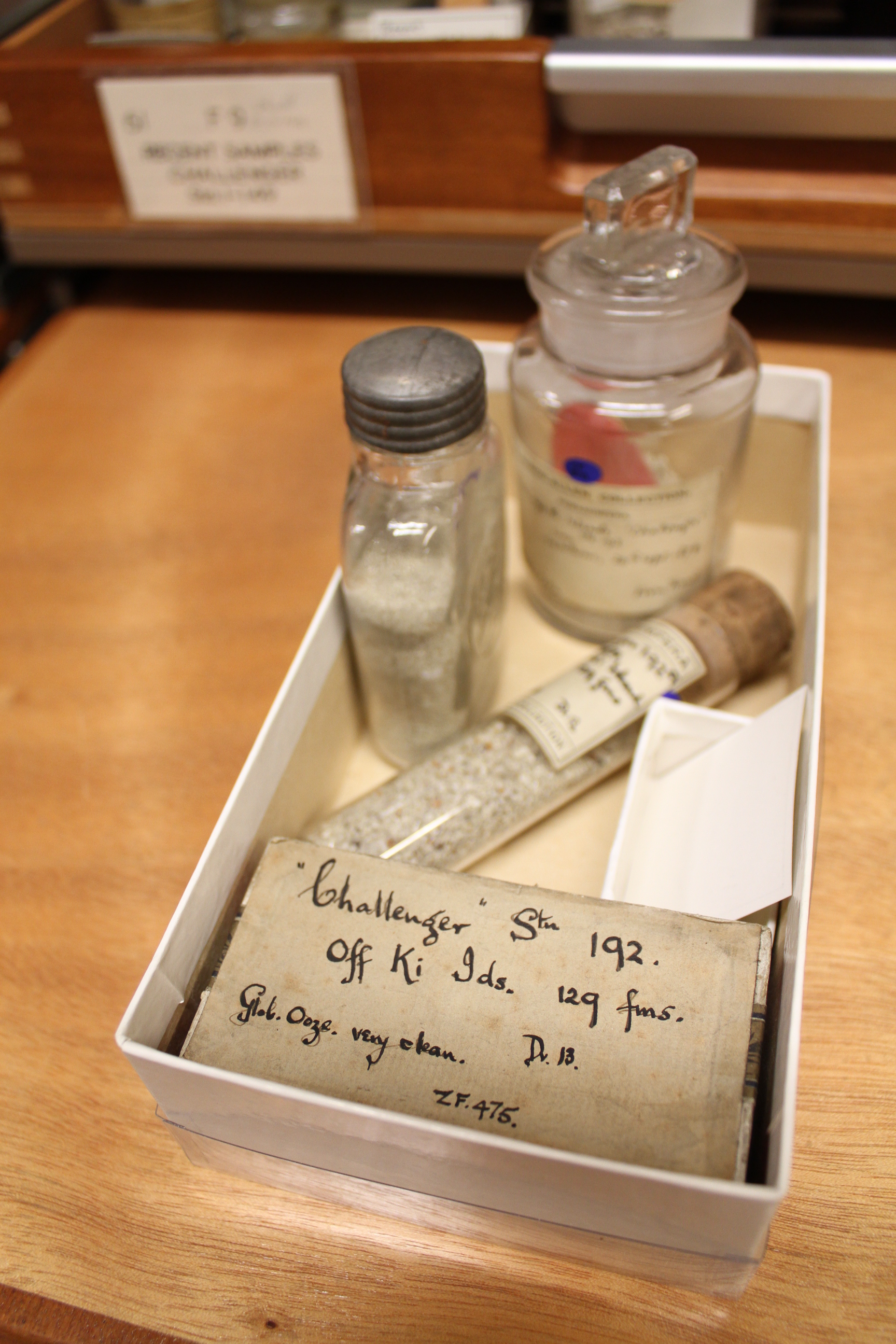
The NHM contains many historical collections such as those from the Challenger Expedition of 1872–76. Image copyright NHM and reproduced with permission.
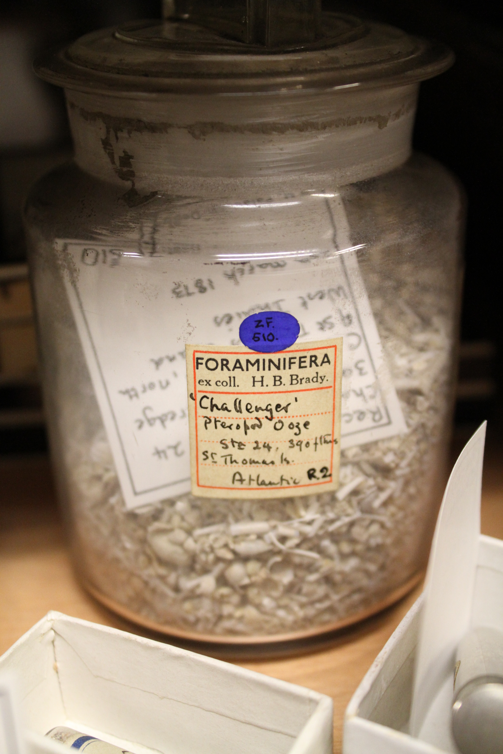
The Challenger Expedition identified over 4,000 previously unknown species, including many pteropods (free-swimming sea snails). Jars as large as this may contain millions of specimens. Image copyright NHM and reproduced with permission.
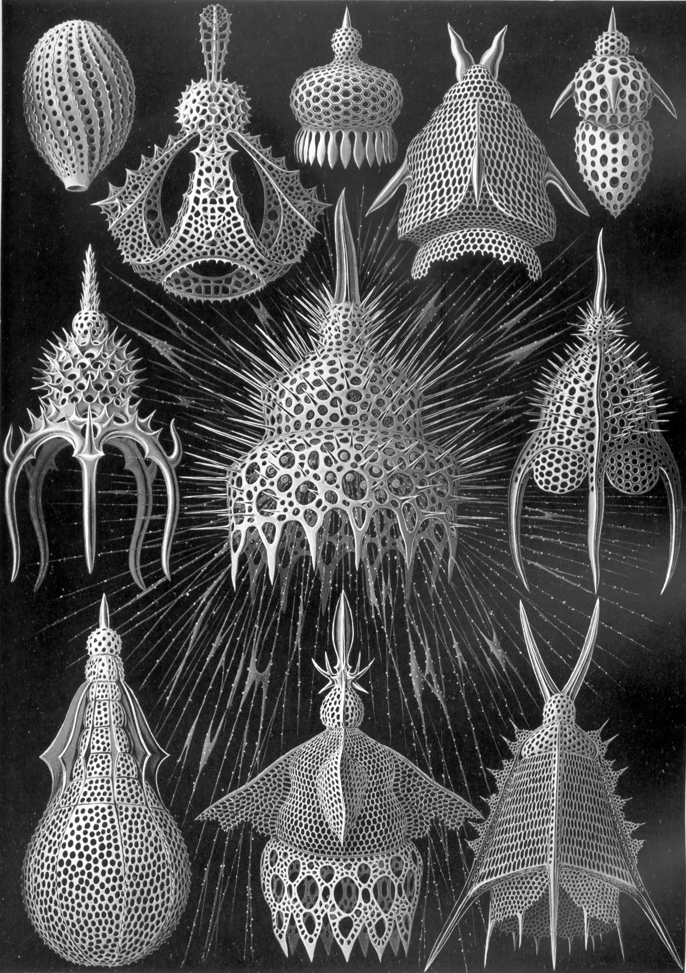
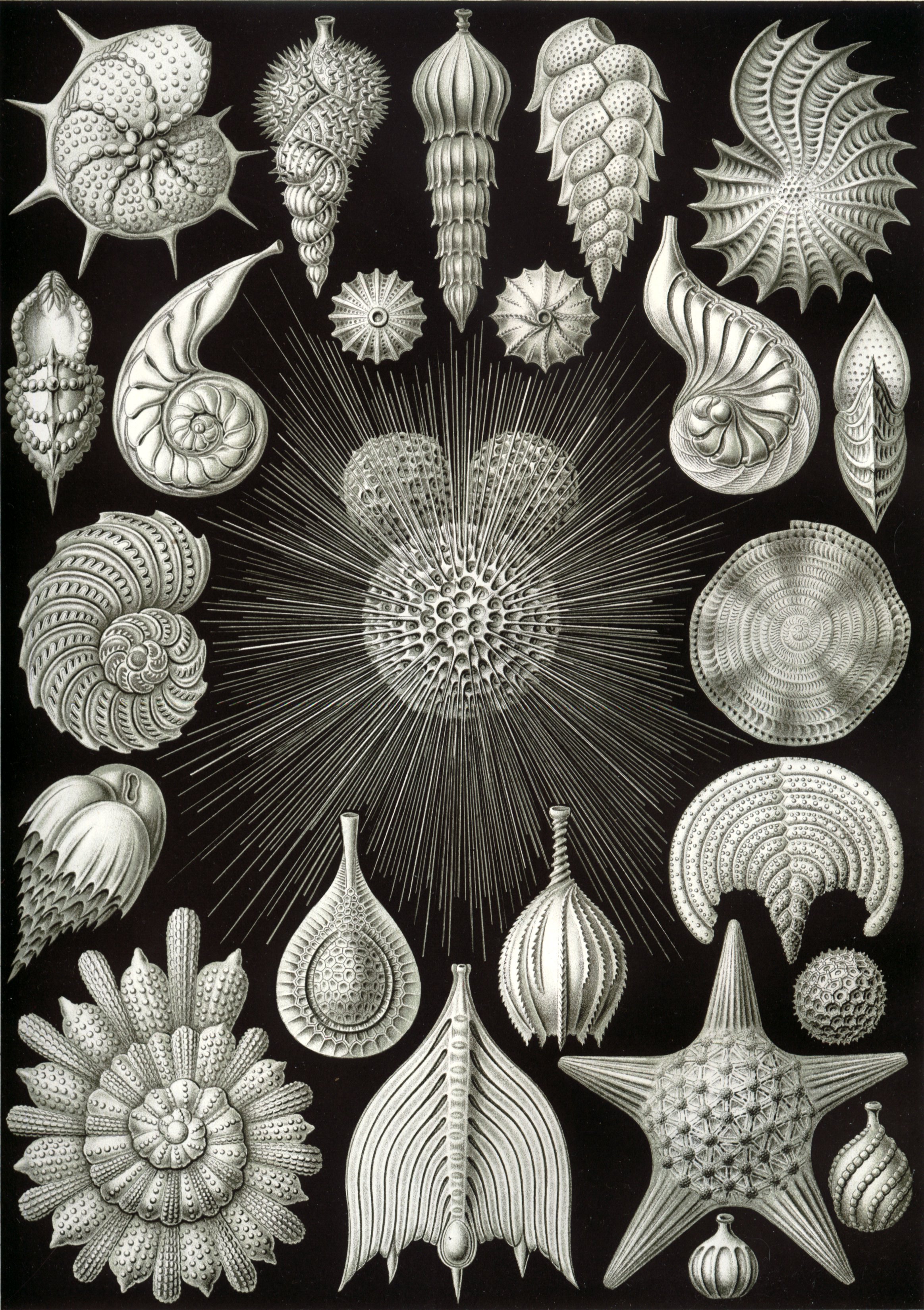
Radiolaria are a group of rhizarians. They were long considered to be single celled animals, but are currently considered to be in a separate kingdom, alongside animals, plants, fungi and others. Many radiolarians were described by Prof. Ernst Haeckel of Jena University from materials collected by the Challenger Expedition. Plate by Ernst Haeckel, Kunst-Formen der Natur, 1899.
Radiolarians produce a skeleton, or test, constructed of silica. These zooplankton live free floating in the water column where they can act as particle feeders, predators or even allow symbiotic algae to photosynthesise for them. Image copyright NHM and reproduced with permission.
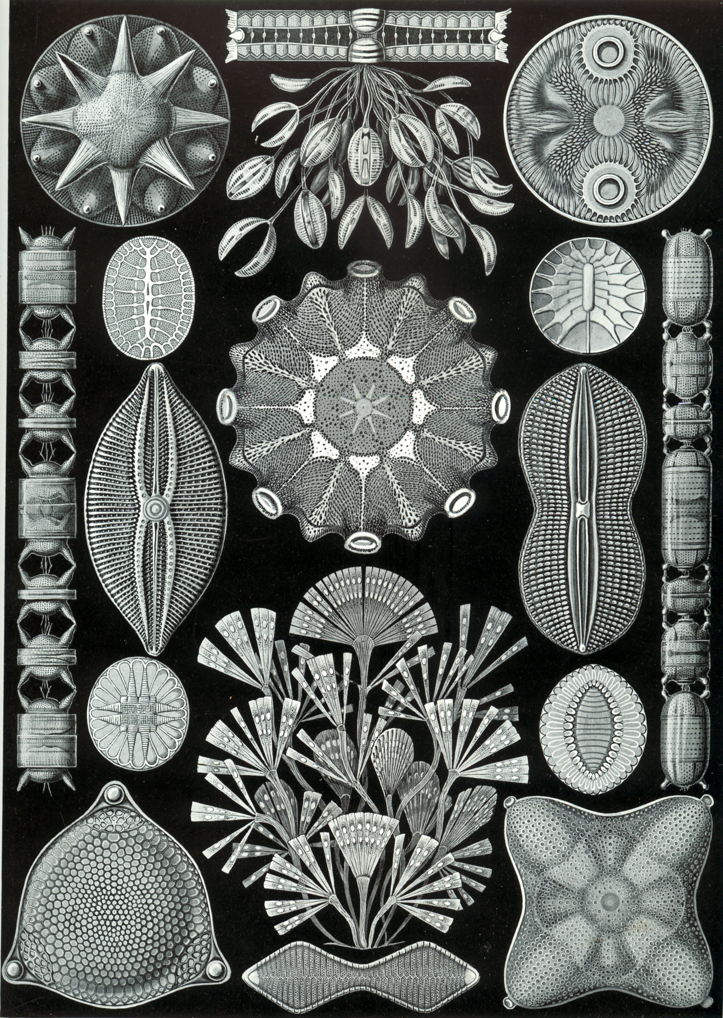
Diatoms are one of the most common groups of phytoplankton today. As radiolarians were once considered to be animals, diatoms were also once considered plants, but are also now considered to be within the kingdom Chromalveolata. Plate by Ernst Haeckel, Kunst-Formen der Natur, 1899.
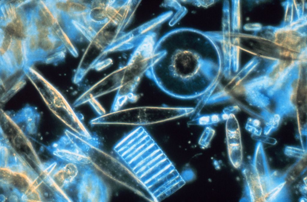
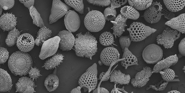
Diatoms produce a silica cell wall termed a frustule. As both diatoms and radiolarians use silica structurally they can become direct competitors for this resource. Image credit: Prof. Gordon T. Taylor, Stony Brook University.
Foraminifera, like radiolarians are rhizarians. They are single celled, but produce multi-chambered tests. Individual tests can reach sizes up to 2cm in diameter. Such tests can commonly be found in the limestone used to build the great pyramids in Egypt. The Egyptians even used these tests as coins. Plate by Ernst Haeckel, Kunst-Formen der Natur, 1899.
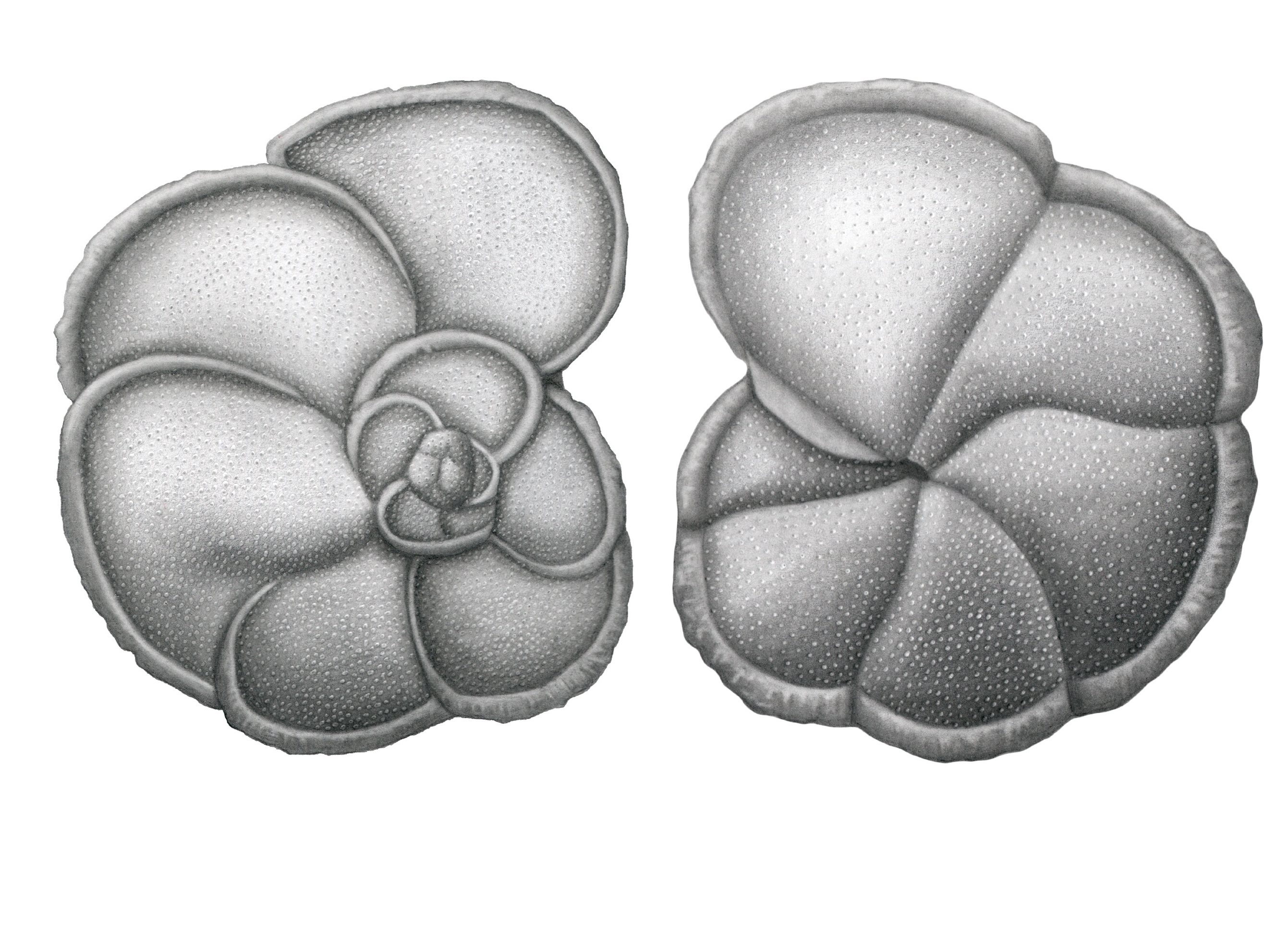
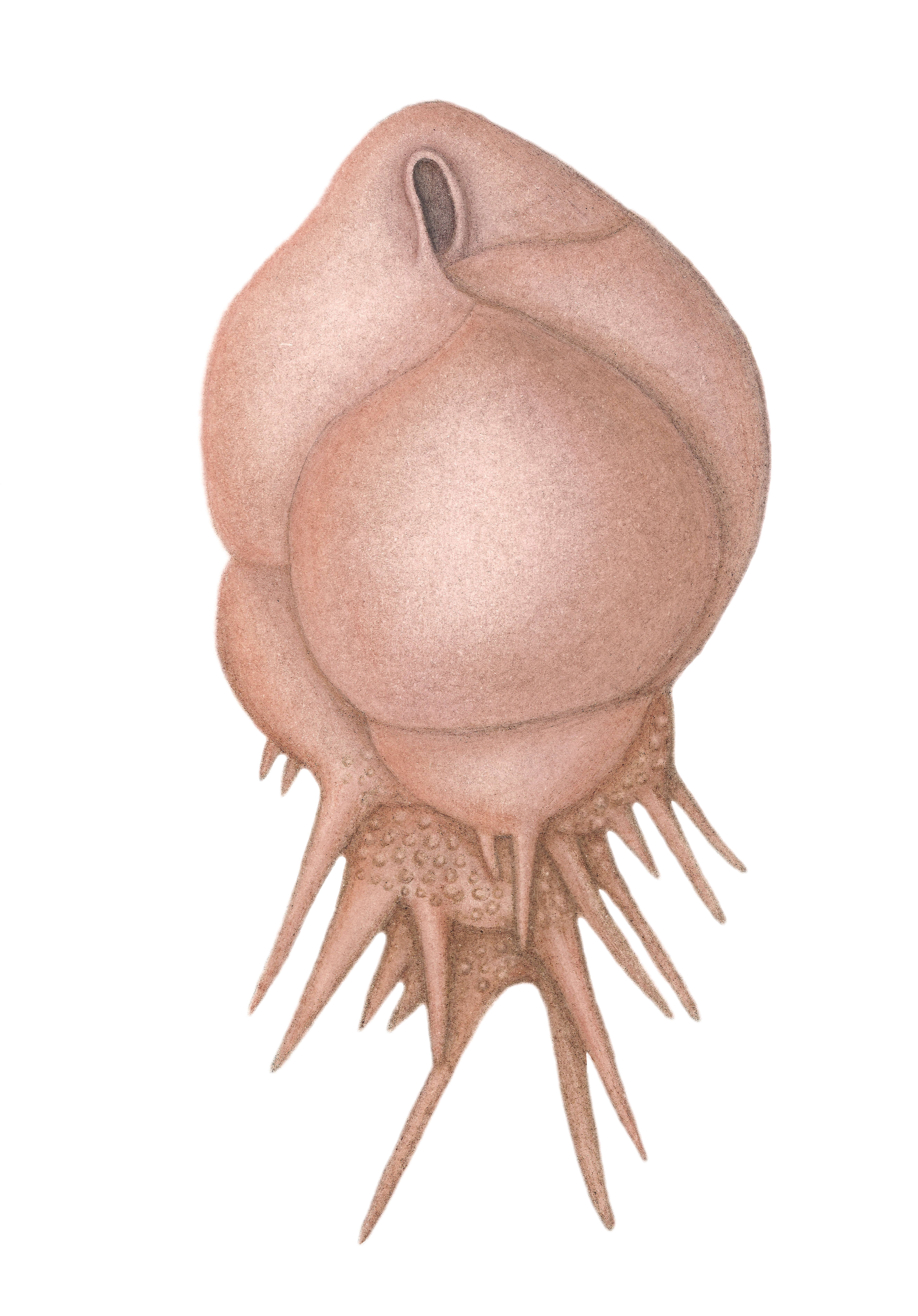
Foraminifera can live either as zooplankton (free floating) or as benthos (on the sea floor) and can produce their tests from a variety of compositions and materials. Agglutinated foraminifera produce their tests using sediment and an organic cement, in a way not too dissimilar to a caddisfly larvae. Many foraminifera, such as planktonic forms, produce their tests of calcium carbonate (CaCO3). The specific chemistry of their tests can be used in inferring palaeoclimates. Pictured: Globorotalia exilis Blow, 1969. Planktonic foraminifera. Stratigraphic range: Middle Pliocene – Early Pleistocene, Geological distribution: Caribbean and Gulf of Mexico regions, low latitudes in open ocean and shallow-water shelf. Hand-drawn reconstruction by Hannah Caine.
This video shows living ostracods; another group of microfossils. Unlike the other groups discussed, ostracods are multicellular animals which belong to the Subphylum Crustacea (best known for crabs, lobsters and barnacles, amongst others). They possess a bivalved carapace (two shells, one on each side) constructed of CaCO3 and are found in a wide range of ecological roles. Video copyright and courtesy of Dr. Dave Horne, Queen Mary University of London and NHM, reproduced with permission.
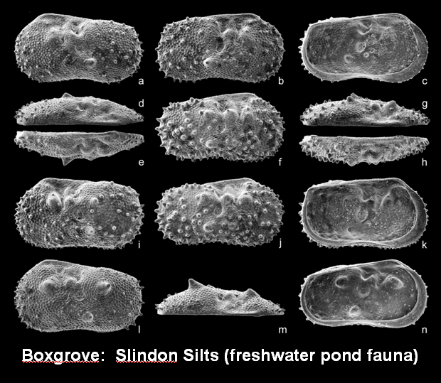
Like a lot of microfossils, ostracods can be restricted to particular environments. In this respect they can be very useful in palaeoenvironmental reconstructions. These freshwater ostracods were discovered in association with some of the first human remains in the UK. The ostracods were used to help interpret the host sediments as a terrestrial deposit with freshwater ponds below a chalk cliff. This kind of information is vital for interpreting archaeological and palaeontological finds. Image copyright NHM and reproduced with permission.
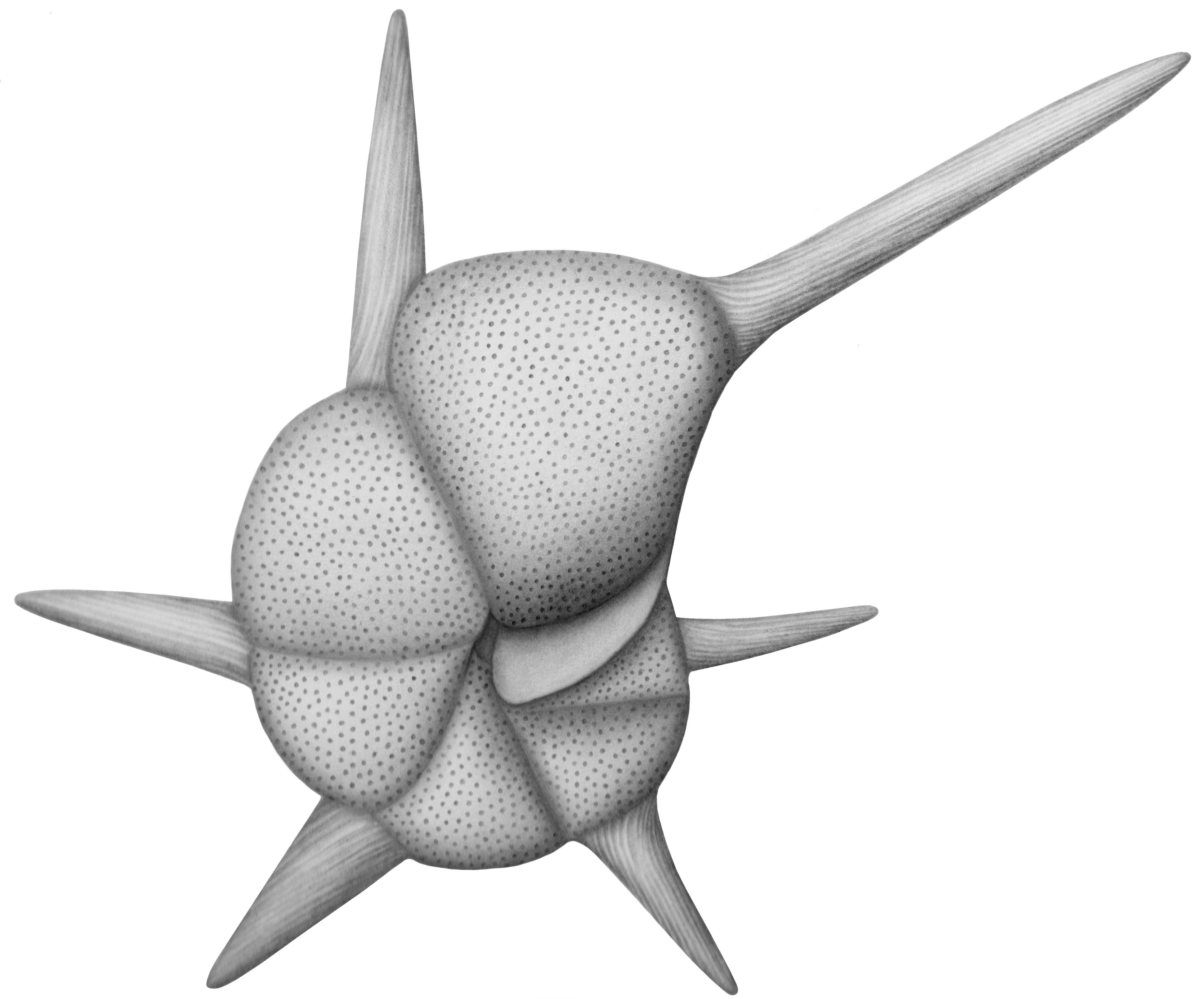
The process of correlating and determining relative ages of rocks based on their constituent fossils is termed biostratigraphy. Microfossils are particularly useful for this as they can be frequently found, widely dispersed and temporally restricted. Pictured: Hantkenina alabamensis Cushman, 1925. Planktonic foraminifera. Stratigraphic range: widely recorded in the late Middle and Late Eocene. Size: maximum diameter (excluding tubulospines) 0.30-60 mm. Geological distribution: Worldwide, mid to low latitudes in open ocean and shallower shelf sites. Hand-drawn reconstruction by Hannah Caine.
The study of microfossils is becoming easier and more accurate with the application of new imaging techniques. High resolution CT scans enables the 3D reconstruction of foraminifera. The video above shows a 3D rendition of Globigerina prasaepis. Such scans will help researchers better understand the true shape of a species, including the interior, without resorting to destructive techniques like thin sectioning. Video copyright NHM and reproduced with permission.
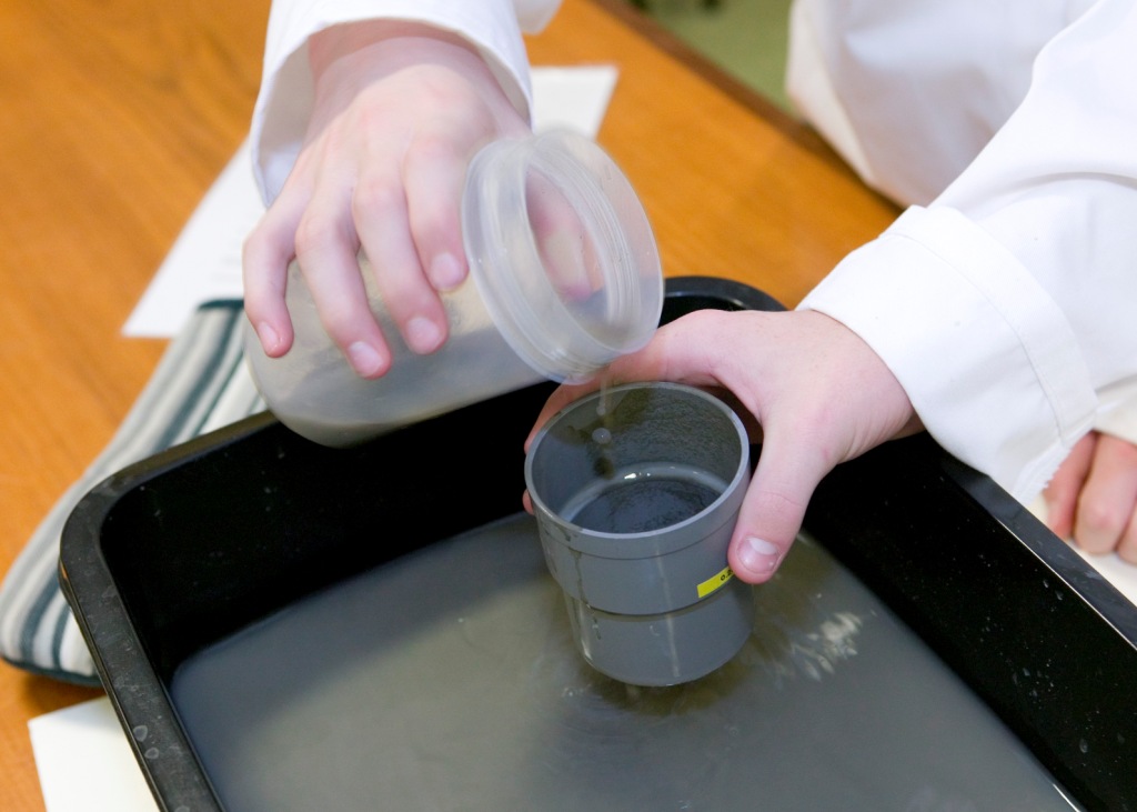
The NHM also uses its collection for teaching science with a hands-on approach. The children perform a biostratigraphical analysis of sediment and determine an age for it. Not only are they surprised by the beauty of the fossils hiding in the sediment, they learn the values of a scientific investigation. Image copyright NHM and reproduced with permission.
Microfossils have also long served as inspiration for many artists and designers. For such biologically simple, generally unicellular, organisms, they possess such intricate, delicate and almost mathematical structures. Pictured: Bulimina aculeata d´Orbigny, 1826. Calcareous benthic foraminifera. Stratigraphic range: Recent. Geological distribution: worldwide, marine, commonly found in the most outer shelf-upper shelf slope environment. Hand-drawn reconstruction by Hannah Caine.
The above video outlines the story of the Blaschka Glass models, including models of radiolarians, currently on display at the NHM. Video copyright NHM and reproduced with permission.
Related Posts
Episode 174: A History of Dinosaurs in 50 Fossils →

Episode 133: Drawing and Painting Dinosaurs →
Episode 115: Diatoms →